OIL MASSAGE TO BABY-KNOW THE FACT,DR.DEV,M.D.,PEDIATRICIAN AND PEDIATRIC PULMONOLOGIST,SAHIBABAD,GHAZIABAD,DELHI NCR

OIL MASSAGE TO BABY-KNOW THE FACT,DR.DEV,M.D.,PEDIATRICIAN AND PEDIATRIC PULMONOLOGIST,SAHIBABAD,GHAZIABAD,DELHI NCR

OIL massage is an important part of giving health to babies.

Oil maasage should be given to all babies starting at the period of newborn.

The best time to start oil massage to baby is ,approximately 10 days after birth when the umbilical stump falls off automatically.

The best time to give massage is before giving bath and it should be 90 minutes before or after the feed.

If the massage is given in close proximity to feed,there is more chance of vomiting.

The massage should be given preferably by mother so that there occurs a good bonding between mother and child.

The best oil for massaging a baby is pure coconut oil and the next best is olive oil.

If possible the massage should be given in open sun light provided there is no cold wind.

Baby should be unclothed fully during massage in warm ambience.

Massage can be given starting from top to bottom or in reverse order.

Lie the baby on his or her back and start massaging with oil from head.oil the scalp hair and massage gently,then start from mid point of forehead and go towards both sides gently.Massage the nose gently because the bone of baby’s nose is soft.DO NOT INSTIL OIL INTO NOSTRILS  OR EARS.

Massage the back of ears then cheeks then armpits.Chest massage should be started from the midline moving toward both sides(periphery)

Massage the tummy in a clockwise movements of your hands very gently.

Arms should be massaged from low to high for 5-6 times.Then both the palms should be massaged.Legs should be massaged from low to high again 5-6 times ,then soles should be massaged.Oil should always be applied in the area of groin .

Now, the baby should lie on the tummy and back massage should be done starting from centre towards both sides.

Oil massage stimulates the different nerves of the baby,increases blood circulation in different parts of body, thereby helps in promoting growth of the baby.It can also detect some hidden injury when baby starts crying in pain which is easily recognised by mother.By increasing bonding with mother, when the baby enjoy the massage,it also helps in overall development of the baby.

Oil massage should preferably be done two times per day and during winter season, it also protects the baby from cold environment.


Five signs of disturbed sleep in children,Dr.Dev,M.D.,Pediatrician and Pediatric Pulmonologist,Sahibabad,Ghaziabad,Delhi NCR

Sleep is very important state of health.

Sleep is a state in which body and mind repares itself.

Adequate sleep is related to time and quality of sleep.

Either less time of sleep or poor quality of sleep ,may affect the body and mind adversely.

Children require more hours of sleep in comparision to adults.

A newborn,less than 4 weeks of age,require 16-18 hours of sleep per day.

A school going children,6-12 years of age require 9-10 hours of sleep per day.

Sleep is essential for the smooth functioning of body and mind.

Following are the 5 signs of disturbed sleep.

1.MORNING HEADACHE:

This is a very important sign of inadequate sleep.It is caused by CO2 retention due to sleep apnea(stoppage of breathing for more than 20 seconds)

2.HYPERACTIVITY:

This is a separate disease entity in children due to many causes,but sleep disturbances is an important cause.

3.ATTENTION DEFICIT:

Excessive sleepiness during day time and poor academic performances is an important sign of inadequate sleep but the child may become inattentive with less capacity to concentrate on anything due to less and bad quality of sleep.

4.RESTLESSNESS AND FRAGMENTED SLEEP:

Frequent arousal during sleep and excessive movement of any part of body is an important sign of disturbed sleep.

5.SOUND DURING SLEEP:

Sleep should ideally be soundless.Any audible sound during sleep is a sign of sleep disordered breathing.

Noisy breathing in the form of snoring is an important sign of upper airway obstruction,which leads to poor growth of body and mind of child and in long term ,it may cause heart disease in children.


FFP- an option to treat Hereditary Angioedema,Dr.Dev,M.D.,Pediatrician and Pediatric Pulmonologist,Sahibabad,Ghaziabad,Delhi NCR

Hereditary angioedema(HAE) is a   rare disorder caused by deficiecny of C1 –inhibitor(C1- INH) since birth.

There are two types of HAE,TYPE 1 and TYPE 2

In type 1 HAE ,there is hereditary deficiency of C1- INH ,whereas in type 2,the level of C1- INH in serum may be normal or elevated but the C 1-INH function is impaired.

C 1- INH interact with approximately 60 other proteins in the body to protect the body from infections.

In its absence,there is failure of degradation of bradykinin, the level of which rises in blood.

Bradykinin,increases the permeability of blood vessels by causing inflammation and dilatation of the vessels.

In effect,the intravascular fluids leak from the blood vessels and accumulate between layers of skin and epithelium.

There occurs swelling all over the body including tongue and lips causing disfigurement.


When the swelling occurs in the respiratory airways ,particularly involving larynx,it may be life threatening,unless treated on emergency basis.

These swellings,usually do not respond to antihistamine,adrenaline,and corticosteroids.

The specific treatment is Berinert , HAEGARDA , or Bradykinin receptor antagonist FIRAZYR(ICATIBANT)which is difficult to get in resource poor countries including India.

There is one case report from Pakistan,a 30 year patient was admitted in emergency after a fall. In the hospital,it was found from the medical record that the patient was a known case of HAE.

The patient got swelling including laryngeal edema which did not respond to antihisamine,adrenaline and corticosterois.The patient developed respiratory distress and went into pulseless cardiac arrest ,revived after CPR and had to be intubated.

Treating doctors decided to give FFP, in view of non availability of specific treatment of HAE.

FFP(Fresh frozen plasma) was given 6 hourly and the patient responded after 18 hours,extubated after 3 days and stepped down from ICU to the ward.He was discharged on danazol prophylaxis and did well on follow up.

There was no adverse effect noted due to FFP.

This is first case report ,published in CUREUS.

FFP contains so many substances apart from C1-INH,which may cause harm to an individual.

If more ,multcentric studies prove its efficacy and safety profile ,,it will be a boon to the patients in resource poor countries.

REFERENCES;OCTOBER 12,2020,In News,By FOREST RAY

Forest Ray received his PhD in systems biology from Columbia University, where he developed tools to match drug side effects to other diseases. He has since worked as a journalist and science writer, covering topics from rare diseases to the intersection between environmental science and social justice. He currently lives in Long Beach, California.
Fact Checked By:
Ines Martins, PhD
Inês holds a PhD in Biomedical Sciences from the University of Lisbon, Portugal, where she specialized in blood vessel biology, blood stem cells, and cancer. Before that, she studied Cell and Molecular Biology at Universidade Nova de Lisboa and worked as a research fellow at Faculdade de Ciências e Tecnologias and Instituto Gulbenkian de Ciência. Inês currently works as a Managing Science Editor, striving to deliver the latest scientific advances to patient communities in a clear and accurate manner.

Corona virus on human skin,Dr.Dev,Pediatrician and Pediatric Pulmonologist,Sahibabad,Ghaziabad,Delhi NCR

It is very difficult to study the virus on human skin due to fear of spread of infection among volunteers.

This is the reason,researchers have chosen to study on the skin of cadaver,which is used for the graft purposes.

Research has proven that the novel corona virus survives on human skin for more than 9 hours.

Influenza A virus survives on human skin for less than 2 hours.

Both viruses are killed when hands are rubbed for 15 seconds using 80% alcohol .

Viruses are also killed when hands are washed with soap water for at least 20 seconds.

Inspite of the fact that novel corona virus spreads mainly by droplets or aerosoles,CDC has recommended sanitizing hands with 60-95% alcohlo based hand sanitizers or washing hands with soap water for at least 20 seconds to check the spread of corona virus.

REFERENCES:https://bit.ly/34vrdlm Clinical Infectious Diseases, online October 3, 2020.

BREATH HOLDING ATTACKS,DR.DEV,M.D.,Pediatrician and Pediatric Pulmonologist,Sahibabad,Ghaziabad,Delhi NCR

Breath holding spells is not uncommon in children.

The phenomenon is very much frightening to the parents.

It is usually seen in children after the age of 6 months.

It usually starts between the age of 6-18 months and mostly observed in the age group of 1-3 years.

It abolishes automatically after 4-6 years of age.

There is no difference between male and female.

There are two types of attacks-1.Pallid spells and 2. cyanotic spells

In pallid spells, the heart rate of the child becomes slow,the child looks pale(more white) and there is loss of consciousness for few seconds ,but definitely ,less than 60 seconds(1 minute).

The pallid spell is usually triggered by pain ,like fall from the bed ,fall on the ground while playing,or minor impact on head of child by gently slapping.

In CYANOTIC SPELLS-

The colour of child becomes blue or purple specially around the lips.It is triggered by aggression,frustation,emotional distrubances and crying in fear or frightening environment to the child.There occurs a short burst of cry or single cry in which at the end of cry the child exhales for a slightly long period and holds the breath in exhalaltion ,not followed immediately by inhalation.There is loss of consciousness for a brief period ,not more than a minute.The muscels of the body may becomes stiff and there may be arching of the body in backward direction.Sometimes,there may be abnormal body movement and rarely there may be seizure.

This phenomena is typically described by parents as the child sometimes forget to take breath.

WHAT TO DO IN THESE SITUATIONS:This condition is harmless to the child with no long term effect on the brain and growth of the child.

The parent should not panic and should be very calm and comfortable.Those objects which may cause injury to the child and particularly sharp objects should be taken away from near of the child.

Make the child lie on the lap.

Do not through water on the face of the child as is usually practiced.It may be dangerous.

The child regain consciousness within a minute.If not,take the child to a doctor/clinic/hospital nearby.

If this episode is the first time,certainly go to the doctor/clinic/hospital.This is because, there may be some serious problems in heart and or brain of the child leading to these symptoms.There may be seizure disorder presenting like this.There may be congenital prolonged QT syndrome presenting like this.

If by proper clinical examination and if required proper investigations,it becomes clear that it is not due to any pathology in the brain and or heart,nothing to be worried.

It has been observed that ,these attacks are more common in children suffering from iron deficiency anemia

It should be investigated and treated ,the child becomes normal.

In some children,the episodes happens many times a day and in others only after few days or months.

It has been seen that ,it happens more frequently in circumstances where parents are more apprehensives.

So,parents should take care of themselves,not to show apprehensions in front of children and not to allow secondary gain to children.

Sometimes ,the episode becomes life threatening,and parents should learn ,how to resuscitate babies by giving mouth to mouth breath and chest compressin if required.

Sometimes ,medications in the form of atropine may be required,which should strictly be used by a qualified medical doctor.

CAUSES OF HAPPY HYPOXIA IN COVID 19,Dr.Dev,M.D.,Pediatrician and Pediatric Pulmonologist,Sahibabad,Ghaziabad,Delhi NCR

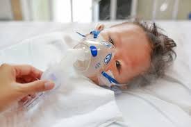
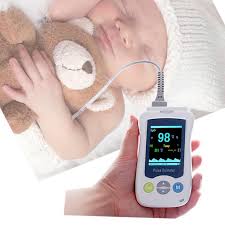
Many patients suffering from COVID 19 are hypoxemic but not in distress and breathing comfortably in room air.This stage is called happy hypoxia.Hpoxemia is detected only after high index of suspician leads to SpO2 monitoring.

Whenever there is less ventilation into the lung,there occurs a  vasoconstriction in the pulmonary vessels as a compensatory response in poorly ventilated area of the lung..This is called hypoxemic Pulmonary vasocontrictor response(HPV).It has been seen that in COVID19 ,this HPV response is diminished with formation of intrapulmonary shunt(right to left) as demontrated by dual energy CT scan. This vasoplegic response is triggered by vasodilator prostaglandin,bradykinin and cytokines.

SARS Cov 2 virus,responsible for COVID 19, damages the endothelium of lung which express angiotensin converting enzyme II receptors, leading to formation of microthrombi in lung capillaries.There is extensive thrombi formation in arteries and capillaries leading to blockage of capillaries and arteries causing ventilation/perfusion mismatch and hypoxemia.This stage is called AVDS(acute vascular distress syndrome)

SARS Cov2 virus damages type 2 pneumocytes and in effect, basement membrane is denued which becomes covered by hyaline membrane consisting of fibrin and dead cells which impaires gas exchange.As the diffusion barrier becomes more pronounced,blood flow through pulmonary vessels increase during exertion.So there is less time for gas exchange during exertion causing hypoxemia.

In normal situations whenever there is less oxygen in the blood,it is sensed by carotid bodies which is situated at the bifurcation of commonl carotid artery..It gives signal to the respiratory centre situated in medulla oblongata which in turn sends signal to diaphragm via phrenic nerve to increase ventilation which is perceived as duspnea by patients.When the PaO2 is between 90 mmHg to 60 mmHg,there is no  such response but as soon as the PaO2 drops below 60 mmHg ,immediately there is a signal exponentially, to increase the ventilation.But for this ventilatory response to occur, when the level of PaO2 is between 40mmHg to 60 mmHg,PaCo2 level must increase to above 39 mmHg to stimulate the carotid bodies.

 SARS COV2 VIRUS BLOCKS the sensitivity of angiotensin converting enzyme 2 receptor present in carotid body .Therefore it can not sense hypoxia or hypercarbia unless it becomes critical.So,there is no signal to increase the ventilation and patients remain comfortable till the hypoxia becomes critical.This stage is called happy hypoxia.

In the early stage of the disease,lung parenchyma is minimally involved which is evident by ground glass opacity in the peripheral regions of  lung.In this stage ,vital capacity is maintained ,there is high compliance and low elastance of lung called L-Phenotype.At this stage there is low  ventilatory response as the hypoxia is vascular in origin due to impairement of HPV  and there is no dyspnea.

As the disease progresses,there is extensive involvement of lung parenchyma with increased ventilatory response,increased permeability of pulmonary capillaries with accumulation of fluid in interstitium(interstitial edema)and inspiratory pressure becomes more negative.This stage is called H-Phenotype with high elastance and low compliance.The transition from L-phenotype to H-Phenotype is precipitaed  by more negative inspiratory pressure created by forceful inspiratory effort by patient during spontaneous breathing,called patient self inflected lung injury(P-SILI)

In case of fever, the oxygen dissociation curve is shifted towards right side, causing low SaO2 for a given PaO2, making the sensing of SpO2 by pulse oxymetry inaccurate as ,at low PaO2 level, SpO2 can underread by about 7 % if SaO2 is below 80%

In case of Diabetes and age more than 65 years the sensitivity of carotid body becomes less causing less ventilatory response.


IN NUTSHELL, at the initial stage of disease,there is minimal involvement of lung parenchyma,primarily the disease affects pulmonary vessels with less  HPV response,AVDS,with development of rifht to left intrapulmonary shunt causing hypoxia,the virus decreases the sensitivity of carotid body to sense hypoxia and increase ventilatory response,shifting of oxygen dissociation curve to right causing low SaO2 for a given PaO2 due to high fever, all leading to less ventilatory response and patients remains comfortable till the critical stage.


REFERENCES:

1. Guan W, Ni Z, Hu Y, et al. Clinical Characteristics of Coronavirus Disease 2019 in China. N Engl J Med. 2020;382(18):1708-1720. doi:10.1056/NEJMoa2002032

2. Mahjoub Y, Rodenstein DO, Jounieaux V. Severe Covid-19 disease: rather AVDS than ARDS? Crit Care. 2020;24(1):327. doi:10.1186/s13054-020-02972-w

3. Lang M, Som A, Mendoza DP, et al. Hypoxaemia related to COVID-19: vascular and perfusion abnormalities on dual-energy CT. Lancet Infect Dis. Published online April 30, 2020. doi:10.1016/S1473-3099(20)30367-4

4. Dhont S, Derom E, Van Braeckel E, Depuydt P, Lambrecht BN. The pathophysiology of ‘happy’ hypoxemia in COVID-19. Respir Res. 2020;21(1):198. doi:10.1186/s12931-020-01462-5

5. Ackermann M, Verleden SE, Kuehnel M, et al. Pulmonary Vascular Endothelialitis, Thrombosis, and Angiogenesis in Covid-19. N Engl J Med. 2020;383(2):120-128. doi:10.1056/NEJMoa2015432

6. Mahjoub Y, Rodenstein DO, Jounieaux V. Severe Covid-19 disease: rather AVDS than ARDS? Crit Care. 2020;24(1):327. doi:10.1186/s13054-020-02972-w

7. Mason RJ. Pathogenesis of COVID-19 from a cell biology perspective. Eur Respir J. 2020;55(4). doi:10.1183/13993003.00607-2020

8. Mo X, Jian W, Su Z, et al. Abnormal pulmonary function in COVID-19 patients at time of hospital discharge. Eur Respir J. 2020;55(6):2001217. doi:10.1183/13993003.01217-2020

9. Hopkins SR. Exercise Induced Arterial Hypoxemia: The role of Ventilation-Perfusion Inequality and Pulmonary Diffusion Limitation. In: Roach RC, Wagner PD, Hackett PH, eds. Hypoxia and Exercise. Advances in Experimental Medicine and Biology. Springer US; 2007:17-30. doi:10.1007/978-0-387-34817-9_3

10. Masi P, Bagate F, d’Humières T, et al. Is hypoxemia explained by intracardiac or intrapulmonary shunt in COVID-19-related acute respiratory distress syndrome? Ann Intensive Care. 2020;10(1):108. doi:10.1186/s13613-020-00726-z

11. Reynolds AS, Lee AG, Renz J, et al. Pulmonary Vascular Dilatation Detected by Automated Transcranial Doppler in COVID-19 Pneumonia. Am J Respir Crit Care Med. Published online August 6, 2020:rccm.202006-2219LE. doi:10.1164/rccm.202006-2219LE

12. Mohan R, Duffin J. The effect of hypoxia on the ventilatory response to carbon dioxide in man. Respir Physiol. 1997;108(2):101-115. doi:10.1016/S0034-5687(97)00024-8

13. Gattinoni L, Chiumello D, Caironi P, et al. COVID-19 pneumonia: different respiratory treatments for different phenotypes? Intensive Care Med. 2020;46(6):1099-1102. doi:10.1007/s00134-020-06033-2

14. Weisbrod CJ, Eastwood PR, O’Driscoll G, Green DJ. Abnormal ventilatory responses to hypoxia in Type 2 diabetes. Diabet Med J Br Diabet Assoc. 2005;22(5):563-568. doi:10.1111/j.1464-5491.2005.01458.x

15. Kronenberg RS, Drage CW. Attenuation of the ventilatory and heart rate responses to hypoxia and hypercapnia with aging in normal men. J Clin Invest. 1973;52(8):1812-1819. doi:10.1172/JCI107363

16. Tobin MJ, Laghi F, Jubran A. Why COVID-19 Silent Hypoxemia Is Baffling to Physicians. Am J Respir Crit Care Med. 2020;202(3):356-360. doi:10.1164/rccm.202006-2157CP

17. Kelman GR. Digital computer subroutine for the conversion of oxygen tension into saturation. J Appl Physiol. 1966;21(4):1375-1376. doi:10.1152/jappl.1966.21.4.1375

WHEN AND HOW TO START BABIES ON SOLID FOODS(WEANING),DR.DEV,M.D.,Pediatrician and Pediatric Pulmonologist,Sahibabad,Ghaziabad,Delhi NCR

It is very common question and very common mistakes by parents,regarding when and how a solid food is introduced to baby.All babies should exclusively be breast fed right from birth ,preferably starting within 30-60 minutes of birth till the age of 6 months.Some mother says that since the child is born through caesarean section(OPERATION),I have started the baby to feed through bottle with formula milk.This is not the correct reason to start formula feeds.Milk production is very less during initial 3 days of child birth but it is sufficient for the baby and it is highly nutrItious and immunobooster to the child.Milk production begins only when the baby starts sucking.If the baby sucks more ,the milk production will be more,if the child sucks less,the milk production will be less and if the child does not suck,there will be no milk production.

Solid foods should be started at the completion of 6 months of age.It should be noted by parents that the child is able to hold his or her head before starting solid foods.

It can be started with any food available in the house but it should be in purees form.There is sufficient evidence from agriculture based families that they start with any grain available to them like corn,millets,rice,wheat.What they do it that,they make the grain in powder form and mix it with animal milk and feed the baby.We have sufficient evidence from hunter and gatherers family that they start giving meat that is made like purees.

So,it is safe to start with any food items like rice,millets,banana,apple,wheat but it should be in the form of purees.It is a good practice that mother should taste the food before feeding the baby.If mother can propel the food placed over tongue through the roof of mouth towards the back of tongue  very easily,it can be given to baby safely.

It should be noted that ,it is the time, just to start the baby on solid foods,not to completely feed the baby with solid foods.The staple food for the baby is still breast milk.So,give the starting food 2-3 times daily and continue one item for 1 week then change to another item.

Some baby may reject the food .In such circumstances,try giving the food with spoon 3 times before stop giving for that session.It has been seen that on third attempt most babies accept the feed.If the baby tries to hold the spoon,let the baby do it,and you can feed them with another spoon.

NOTE:The baby  should be seated upright as shown in picture at the outset, and food should not be sticky or lumpy to avoid the chance of chocking.

SPICY AND FLAVOURED FOOD:These food items can be given at this age of weaning the child.Since the child is already exposed to these spices and flavours in food through breast milk or even before birth in the uterus through the blood of mother,the child accepts it and there is no harm to the child.

ALLERGIC FOODS:Allergic food items can safely be started at this age and moreover,introducing allergic foods like egg,peanuts,fishes at this age may lessen the chance of developing allergy later in life.

NOTE:If the elder sibling is allergic to some food item,it is mandatory to get the baby tested for allergy before giving that food.

Food items should be changed every week and all kinds of foods should be introduced by the age of 9 months.It has been observed that by doing so,the child accepts all kinds of food items later in life.

Breast feed should be continued till the age of 2-3 years.It has been observed that the child who continue to breast feed till 2-3 years ,accepts all home made foods easily in comparision to those who continue breast feed for lesser period.

The quantity of solid foods should be increased gradually and by the age of 9 months, some lumpy foods which dissolves easily in mouth or mashed foods should be given to baby.Teeth are not erupted completely at this age but the child learns to manage food items with gums and propel towards back of tongue.By doing so ,the chewing muscles also gain strength.

NOTE;There should not be particles of food in mouth after getting broken in mouth by baby to avoid choking.

Honey should not be given before 1 year of age due to risk of BOTULISM

It is a common practice in India to start weaning with DAL WATER(Pulse water) It is a wrong practice.Start with whole DAL in the form of purees,(any dal)then give khichri after 1 week,then give rice and pulse after 1 week like that.

you can make powder of multiple grains and give one grain mixed with breast milk or animal milk at a time then add another after 1 week and gradually give multiple grains after 4-5 weeks . Fruits and vegetables should be given in the same manner.

REFERENCES:

Abrams EM and Becker AB. 2015. Food introduction and allergy prevention in infants. CMAJ. 187(17):1297-301.

American Academy of Pediatrics. 2017. Starting Solid Foods. Retrieved from https://www.healthychildren.org/English/ages-stages/baby/feeding-nutrition/Pages/Switching-To-Solid-Foods.aspx (12/28/2018).

American Academy of Pediatrics Committee on Nutrition. 2000. Hypoallergenic infant formulas. Pediatrics. 106(2 Pt 1):346-9.

Cameron SL, Taylor RW, Heath AL. 2015. Development and pilot testing of Baby-Led Introduction to Solids–a version of Baby-Led Weaning modified to address concerns about iron deficiency, growth faltering and choking. BMC Pediatr. 15:99.

Coulthard H, Harris G, Emmett P. 2009. Delayed introduction of lumpy foods to children during the complementary feeding period affects child’s food acceptance and feeding at 7 years of age. Matern Child Nutr. 5(1):75-85.

Daniels L, Heath AL, Williams SM, Cameron SL, Fleming EA, Taylor BJ, Wheeler BJ, Gibson RS, Taylor RW. 2015. Baby-Led Introduction to SolidS (BLISS) study: a randomised controlled trial of a baby-led approach to complementary feeding. BMC Pediatr. 15:179.

de Lauzon-Guillain B, Jones L, Oliveira A, Moschonis G, Betoko A, Lopes C, Moreira P, Manios Y, Papadopoulos NG, Emmett P, Charles MA. 2013. The influence of early feeding practices on fruit and vegetable intake among preschool children in 4 European birth cohorts. Am J Clin Nutr. 98(3):804-12.

Dogan E, Yilmaz G, Caylan N, Turgut M, Gokcay G, Oguz MM. 2018. Baby-led complementary feeding: Randomized controlled study. Pediatr Int. 60(12):1073-1080.

Du Toit G, Foong RM, and Lack G. 2016. Prevention of food allergy – Early dietary interventions. Allergol Int. 65(4):370-377.

Forestell CA. 2017. Flavor Perception and Preference Development in Human Infants. Ann Nutr Metab. 70 Suppl 3:17-25.

Forestell CA and Mennella JA. 2007. Early determinants of fruit and vegetable acceptance. Pediatrics 120(6):1247-1254.

Forestell CA and Mennella JA. 2017. The Relationship between Infant Facial Expressions and Food Acceptance. Curr Nutr Rep. 6(2):141-147.

Harris G and Mason S. 2017. Are There Sensitive Periods for Food Acceptance in Infancy? Curr Nutr Rep. 6(2):190-196.

Howcroft R. 2013. Weaned Upon A Time: Studies of the infant diet in prehistory. Stockholm.

Ierodiakonou D, Garcia-Larsen V, Logan A, Groome A, Cunha S, Chivinge J, Robinson Z, Geoghegan N, Jarrold K, Reeves T, Tagiyeva-Milne N, Nurmatov U, Trivella M, Leonardi-Bee J, Boyle RJ. 2016. Timing of Allergenic Food Introduction to the Infant Diet and Risk of Allergic or Autoimmune Disease: A Systematic Review and Meta-analysis. JAMA. 316(11):1181-1192

Liem JJ, Huq S, Kozyrskyj AL, Becker AB. 2008. Should Younger Siblings of Peanut-Allergic Children Be Assessed by an Allergist before Being Fed Peanut? Allergy Asthma Clin Immunol. 4(4):144-9.

Mennella JA, Reiter AR, Daniels LM. 2016. Vegetable and Fruit Acceptance during Infancy: Impact of Ontogeny, Genetics, and Early Experiences. Adv Nutr. 7(1):211S-219S.

Mura Paroche M, Caton SJ, Vereijken CMJL, Weenen H, Houston-Price C. 2017. How Infants and Young Children Learn About Food: A Systematic Review. Front Psychol. 8:1046.

Okubo H, Miyake Y, Sasaki S, Tanaka K, Hirota Y. 2016. Feeding practices in early life and later intake of fruit and vegetables among Japanese toddlers: the Osaka Maternal and Child Health Study. Public Health Nutr. 19(4):650-7.

West C. 2017. Introduction of Complementary Foods to Infants. Ann Nutr Metab. 70 Suppl 2:47-54.

title image of baby touching hands in high chair by lmnop88a / flickr

imagine of baby foods by Frédérique Voisin-Demery / flickr

image of baby making funny face while eating by Fimb /flickr

image of baby turning away from food by Abigail Batchelder /flickr

image of baby grabbing spoon by César Rincón / flickr

Content last modified 1/2019


VACCINATION(IMMUNISATION),IN CHILDREN,Dr.Dev,M.D.,Pediatrician and Pediatric Pulmonologist,Sahibabad,Ghaziabad,Delhi NCR

Vaccination(Immunisation) is a process  in which a modified part of disease producing organism is given into the body, either through mouth or through injections.This modified part is not capable of causing disease but capable of activating the body to produce a substance in the body which kills the disease producing organism which subsequently invades the body.This is called active immunisation.It is done before any organism attacks the body.

When an organism has already attacked the body and body is not capable of fighting it,antibody already available is injected into the body to kill that organism and it is called passive immunisation

Immunisation starts from birth, when no organism has yet attacked the body. When  it is not done ,antibody which has come from mother to newborn acts as a killer to any attacking organism,provided it is already present in mother.

SCHEDULE OF VACCINATION:

AT BIRTH(1.);BCG-to protect against Tb(Tuberculosis) is given on left arm and a scar may form at the site after healing of the wound formed 4-6 weeks after the vaccination.Scar persists lifelong.

(2) Hepatitis B first dose is given to protect against hepatitis ,a liver disease.

(3)Oral polio vaccine is given through mouth to protect against polio disease.

AT 6 WEEKS OF AGE:a combination vaccine is given which contains DPT,IPV,Hepatitis B,Hemophilus influenzae b. DPT protects against Diphtheria,Tetanus and Pertussis.IPV is injectable polio vaccine to protect against polio,Hemophilus vaccine protects against a bacterial infections causing pneumonia and meningitis.

At this age Pneumococcal conjugate vaccine can be given to protect against severe form of pneumonia and Rotavirus vaccine can be given orally to protect against severe form of diarrhoea.

AT 10 WEEKS OF AGE: All vaccines given at the age of 6 weeks are repeated.

AT 14 WEEKS OF AGE:All vaccines given at the age of 10 weeks are repeated except Rotavirus if R1 has been used.If R5 has been used for Rota virus protection,then it is also repeated as third dose.

AT 6 MONTHS OF AGE;The first dose of influenza vaccine is given to protect against influenza virus causing severe form of pneumonia in children.

AT THE AGE OF 7 MONTHS;The second dose of influenza vaccine is given which is then repeated every year.

AT 9 MONTHS OF AGE;MMR vaccine is given to protect against viral diseases caused by measles,mumps,and rubella viruses.

At this age Typhoid conjugate vaccine can be given as a single dose to protect against Typhoid fever and Meningococcal conjugate vaccine can be given to protect against meningitis(brain fever) caused by Meningococcus.It is repeated after 3 months,.

AT 1 YEAR OF AGE;Hapatitis A vaccine is given to protect against hepatitis ,liver disease.At this age ,Encephalitis vaccine(JEEV) can be given to protect against Japanese encephalitis ,a lethal disease.This vaccine(JEEV) is repeated after 4 weeks.

AT THE AGE OF 15 MONTHS:MMR vaccine is repeated .At this age, Varicella vaccine is given to protect against chicken pox.The booster dose of Pneumococcal conjugate vaccine is given at this age.

AT THE AGE OF 18 MONTHS:BOOSTER doses of DPT,IPV and Hepatitis A  is given.Booster dose of Hemophilus influenza type b is also given at this age.

AT THE AGE OF 2 YEARS; Vi Polysaccaride vaccine may be given to protect against Typhoid to those child who has not received Typhoid conjugate vaccine

AT THE AGE OF 4-6 YEARS;BOOSTER DOSES of DPT,MMR AND VARICELLA are given at this age.

AT THE AGE OF 10 YEARS;TdaP Vaccine is now recommended which acts as a booster for Diphtheria,Tetanus and Pertussis.For girl child ,HPV vaccine is given to protect against cervical cancer and it should be repeated after 1 -2 months and 6 months of first dose.

AT THE AGE 16 YEARS:TdaP vaccine is repeated.


POST EXPOSURE VACCINE;In case of dog bite or bite from any wild or unknown animal ,a vaccine is given to protect against a lethal disease,Rabies.It is 100% lethal disease for which no treatment is available.

Five doses are given on days 0,3,7,14,28.In third degree, bite immunoglobulin is also given as early as possible and not beyond 7 days.

NOTES;a minimum of 4 weeks interval is must between two doses of same vaccine.But, it can be given if one has missed the schedule date.It is safe to be given between 4-8 weeks.

If a child is completely vaccinated, there is no need of repeated Tetanus vaccine after minor injuries.

No vaccine gives 100% protection.

Adverse effects can happen to any vaccine ,most commonly within 30 minutes after vaccination but may occur later on.


;

SISUNATOVIR,A PROMISING DRUG TO TREAT SEVERE RSV INFECTIONS INCLUDING BRONCHIOLITIS,Dr.D.K.JHA,M.D.,Pediatric Pulmonologist,Delhi,India

Bronchiolitis in severe form

RSV(Respiratory syncytial virus) is the most common cause of bronchiolitis in children.

Bronchiolitis is usually mild but in infants between 3-6 months,it may become serious and sometimes life threatening.

It usually becomes serious in preterm infants and infants already having cardiac disease with significant shunt,infants having CLD(Chronic lung disease,previously known as BPD) and infants with primary immune deficiency.

There is no specific treatment other than Rivaverin which may or may not work.

So, the mainstay of treatment is only supportive and it has high morbidity and mortality.

In such situations,a new drug Sisunatovir may be life saving for many infants.

In a research ,66 adults were inoculated with RSV,then they were given treatment with this new drug Sisunatovir.It resulted in significant reduction in clinical symptoms with significant reduction in viral load,with no significant adverse effects.The new drug was well tolerated and there was no resistane to this new drug.It was Phase 2a study.

Sisunatovir binds to the surface protein F, of RSV and inhibits its replication.It is a fusion inhibitor which is administered orally.

REFERENCES:ReViral announces FDA Fast Track designation granted to sisunatovir for the treatment of serious respiratory syncytial virus infection. https://www.businesswire.com/news/home/20200804005065/en/ReViral-Announces-FDA-Fast-Track-Designation-Granted. Accessed August 4, 2020

Meaning of ground glass opacity on Chest CT,Dr.D.K.Jha,M.D.,Pediatric Pulmonologist,Delhi,India

Computed tomography of chest(Chest CT) has become an essential imaging modality to diagnose many chest conditions including some complicated Pneumonia.

As CT chest is time consuming,so children below 3 years should better be sedated for proper examination of chest by CT.

Ground glass opacity (GGO) may be seen in different chest conditions and its location may give some clue to the diagnosis.

In the time of widespread occurance of COVID 19 ,CT chest may be useful to point towards diagnosis when the serological tests are negative.

In COVID 19 Pneumonia typical findings on chest CT is bilateral ground glass opacity located peripherally in the subpleural regions and at lower lobes of lung.Later on there may be crazy pavy changes with architectural damage ,perilobular opacities superimposed upon ground glass lesions.

There may be consolidation in the lower lobes of the lung located in subpleural regions suggestive of COVID19 Pneumonia.

Atypical findings in COVID19Pneumonia may be ground glass opacity on the upper lobes of lung,peribronchovascular regions and,there may be cavitation,lymphadenopathy and pleural thickening.

Ground glass opacities are also seen in other viral pneumonia.It is seen in upto 75%cases of Adenovirus Pneumonia and more than 75%cases of cytomegalovirus(CMV) Pneumonia.It is also seen in approximately 25% cases of Herpes simplex Pneumonia.

It is also seen in Pneumonia due to Pneumocystis carini but in this case it is mainly located on the upper lobes.

GGO may be observed in interstitial lung diseases(ILD) but the pattern of distribution may differentiate it from infective origin.

GGO may be seen in lung injury caused by electronic cigarrette smokes,eosinophilic pneumonia,hypersensitivity Pneumonia,Pulmonary alveolar proteinosis,diffuse alveolar hemorrhage,and pulmonary edema.

Bacterial Pneumonia can be differentiated by the distribution of opacities in the focal ,segmental and lobar regions not predominantly in the lower lobes.Other findings also differentiate it from viral pneumonia, like cavitation,lymphadenopathy and lung abscess

REFERENCES;Cite this: Broad Differential Diagnosis of Chest CT Ground-Glass Opacities – Medscape – Jul 16, 2020.