The most common imaging technique to diagnose a chest contion is Chest X-Ray.
Chest X-Ray has low sensitivity to diagnose a lesion of chest ,particulalry parenchyma of lungs.
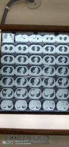
CT Chest ,although gives a high dose of radiation which is harmful particularly for a growing lung of child,is sometimes required to diagnose a chest condtion with more accuracy.
There are so many defined opacities seen on CECT or HRCT chest which gives diagnostic clues.
Ground glass opacities are one of them.
In the time of many cases of Pneumonia due to Corona Virus Disease 2019,it has been observed that the typical findings seen on CECT/HRCT Chest is ground glass opacities(GGO) seen in the periphery of lungs at subpleural locations and in the lower lobes of lungs bilaterally. Later on there may be crazy-pavy pattern,architectural distortion and perilobular opacities superimposed on GGO.There may be bilateral subpleural and lower lobe consolidation. .Atypically, a patient of COVID 19 pneumonia may have upper lobe and peribronchovascular distribution of GGO,cavitations ,pleural thickening, and lymphadenopathy.
It is very dificult to differentiate these lesions typical of COVID19,from other viral pneumonia as approximately 75% of Adenovirus pneumonia and more than 75% of cytomegalovirus and Herpes simplex virus Pneumonia have GGO on chest CT.Approximately 25% of Pneumonia due to Human Metapneumovirus has GGO on chest CT.
ILD(Interstitial lung disease) also shows GGO on chest CT.GGO is commonly seen in Pneumocystis carini Pneumonia but in such cases it is predominantly seen on upper lobes.
GGO is commonly seen in eosinophilic pneumonia,pulmonary edema,alveolar hemorrhage,hypersesitivity pneumonitis ,pulmanary alveolar proteinisis and lung injury due to vaping and use of electronic cigarettes.
These may be differentiated by clinical pictures.
Bacterial pneumonia may be differentiated from viral Pneumonia as the opacity has focal lobar,segmental,and sunsegmental distribution usually not predominantly in the lower lobes..It may be further differentiated by the presence of cavity.lung abscess and lymphadenopathy
REFERENCES:https://bit.ly/3exnOFJ Radiology, online July 7, 2020.