Dr.Dev Kumar Jha
Alumnus of AIIMS New Delhi
Founder Director-Vitalis Super Speciality Children Hospital ,D64/1-Dilshad Colony,New Delhi 110095
Dr.Dev Kumar Jha
Alumnus of AIIMS New Delhi
Founder Director-Vitalis Super Speciality Children Hospital ,D64/1-Dilshad Colony,New Delhi 110095
ABSTRACT:
Tropical Pulmonary eosinophilia is a rare disease affecting multiple organs but there is more involvement of lungs.It ia caused by hypersensitivity reactions to trapped microfilariae of Wuchereria bancrofti(W.bancrofti) and Brugia malayi(B.malayi) in the lungs. I report a case of TPE(Tropical Pulmonary eosinophilia in a child who presented similar to Pulmonary tuberculosis,diagnosed and treated timely with good response.
KEYWORDS Tropical pulmonary eosinophilia,hypersesitivity reactions, mocrofilaria,Filaria
INTRODUCTION:
Tropical Pulmonary eosinophilia as a disease entity was first described in 1940 and the term Pseudotuberculosis with eosinophilia was given.In 1943 Weingarten coined the term TPE(Tropical Pulmonary eosinophilia) for this disease. The disease occurs in costal India like Maharashtra,Kerala,Goa and odisha.It has also been found in some areas of Bihar( prevalence of 9% in jail inmates of Patna).A prevalence of 0.5% among children of Tamil Nadu has been found.It occurs in filaria endemic zones of South America,Africa and South East Asia.The common presentation is cough,difficulty in breathing,fever and weight loss. On investigation there is Leucocytosis with eosinophilia,raised serum IgE and raised specific serum IgE and IgG against Filaria antigen. Treatment is 3-4 weeks course of DEC(Diethylecarbamzepine)
CASE SUMMARY:
9 years old female child came to my OPD with complaint of fever off and on for 2-3 months,cough for 2 months,difficulty in breathing for 1 month ,breathless on exertion for 1 month.
There was no contact history for tuberculosis in the family or relatives
She was taking treatment from different doctors in past 3 months
She was also loosing her weight in last 2 months and she was easily fatigued in last 1 month
on clinical examination she was underweight and anemic,there was significant cervical lymphadenopathy ,no clubbing,no cyanosis and on chest auscultation there were bilateral wheezes and crackles.Abdominal examination showed hepatsplenomegaly
On investigation
Hb 14.3g/dl,
TLC-50,000/cmm,
Neutrophil 28%,lymphocytes10%,
Eosinophil 61%
Basophil 0%,ESR18mm/1H,
Peripheral smear examination-leucocytes increased in number with normal distribution, platelets adequate,no blast cells, malaria parasites amd microfilariae absent
Chest X-Ray-multiple bilateral miliary mottling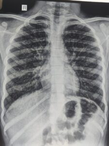
I thought it may be a case of
1.Tuberculosis with some parasitic infections,
2.Asthma with ABPA,
3.hypereosinophilic syndrome,
4.Tropical Pulmonary Eosinophilia
Then , I investigated further
GA for CBNAAT -Negative,Montoux test -negative
Serum IgE -3389U/L,Serum specific filaria antibody IgE and Ig G were raised
Then,I Put the child on DEC 5 mg/kg/day for 4 weeks orally and child started improving after 1 week of initiating DEC
Repeat Serum IgE after 4 weeks was normal ,TLC came down to normal range, and repeat chest X-Ray at that time was normal,child started gaining weight
DISCUSSION:
TPE is usually seen between the age group of 15-45 years .It is rare to see in children less than 15 years ,which was our case. The male:female is 4;1 which is against our case.The clinical features are malaise,fatigue,cough ,chest pain, breathlessness which are seen more during night.Clinical findings in chest may be wheezes and crackes but in 20% cases,there may be no findings in chest.It may involve any system apart from chest leading to the findings of cervical lymphadenopathy,hepatomegaly,splenomegaly,hepatosplenomegaly
The pathology involves hypersensitivity reactions type I,III,and IV to microfilariae.It is due to release of microfilariae from lymphatics which get entrapped into pulmonary circulation.Degranulation of eosinophils, release major basic proteins along with other granules which destruct microfilariae as well as respiratory and terminal bronchioles with lung parenchyma.In long standing untreated cases fibrosis develops.
Lung function test shows mixed obstructive and restrictive pattern of lung disease.
Chest radiograph shows raticuonodular opacities in mid zone and lower zone but as the disease progresses,there are miliary mottling all over the lung fields. Chest X-Ray may be normal in about 20% cases.CT chest may show mediastinal lymphadenopathy,consolidation,bronchiectasis,cavitation and Pleural effision.
CBC shows leococytosis with marked eosinophilia,AEC is usually more than 3000/cmm,Total serum IgE and specific IgE and IgG against Filaria is raised.
ELISA test detecting 0g4c3 antigen is sensitive and specific to W.bancrofti and Sanwich ELISA detecting recombinant antigen Bm-SXP-1 is specific to B.malayi
Trearment is DEC,5mg/kg/day for 4 weeks.Steroids may have a role but dose and duration is yet to be known
Our case is well fitting into the diagnostic criteria
CONCLUSION: TPE is a hypersensitive reaction to W.Bancrofti and B.malayi microfilariae mainly type I, causing eosinophilic alveolitis with inflammation of terminal an d respiratory bronchioles.Diagnosed by clinical features ,mainly night symptoms of cough and dyspnea,peripheral eosinophilia which has diurnal variation and paradoxically low level during night,raised serum IgE and specific IgE and IgG against filaria ,infiltrates on chest radiograph.Treatment is oral DEC for 4 weeks but there is 20% relapse within 5 years. The disease closely resembles Pulmonary tuberculosis and asthma.Untreated and partialy treated case may progress to Interstitial lung disease(ILD)
REFERENCES:
1.Indian J Med Res. 2013 Sep; 138(3): 295–302.
2. Udwadia FE. Herzog H, editor. Tropical eosinophilia. Pulmonary eosinophilia: progress in pulmonary research. 1975;VII:35–155. Basel: S Karger
3.Neva FA, Kaplan AP, Pacheco G, Gray L, Danaraj TJ. Tropical eosinophilia. A human model of parasitic immunopathology, with observations on serum IgE levels before and after treatment. J Allergy Clin Immunol. 1975;55:422–9
4. Frimodt-Moller C, Barton RM. A pseudo-tuberculosis condition associated with eosinophilia. Indian Med Gaz. 1940;75:607–13
ABSTRACT;
Necrotising pneumonia is a serious and uncommon complication of pneumonia in children.It usually occurs in previously healthy children.It should be suspected when a child with pneumonia worsens clinically after 72 hours of appropriate antibiotics. It may be complicated by pyopneumothorax and Broncopleural Fistula. It is diagnosed by radioimaging of chest and CECT Chest is the best modality.It needs long term antibiotics with long hospital stay.
CASE SUMMARY:
8 years male child came in our emergency with history of fever for 4 days with weakness and breathing difficulty for 2 days
On reaching hospital ,in the emergency department the child lost his consciousness
On examination ,the child was in shock and breath sound was diminished on right side of chest
The child had to put on ventilator immediately and then chest X-Ray was done which revealed right sided pneumothorax
Chest tube drainage was done immediately,which relieved pneumothorax
Appropriate antibiotics were started but the condition of the child did not improve
Pus started coming out in the chest tube
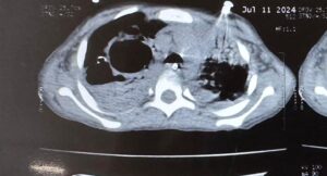
CECT Chest was done which revealed cavitary lesions (thin walled cavities) with distortion of lung architecture with pyopneumothorax
There were appearance of air bubble in the chest tube which was not earlier after relieving of pneumothorax
It was then confirmed that there has been formation of Bronchopleural Fistula(BPF)
In view of necrotising Pneumonia and BPF, antibiotics was upgraded to clindamycin and Meropenem
Ventilator settings was adjusted with low Ti. Low PEEP and minimum Tidal Volume,

 en
en
General condition improved gradually and child became afebrile,OG tube feeding was started while on ventilator
Condition became permissible to extubate the child after 2 weeks of mechanical ventilation
While on ventilator ,the child had persistent metabolic alkalosis after the first report on ABG of respiratory acidosis before ventilation,there was persistent hypokalemia ,then one episode of respiratory alkalosis,all were managed properly. Child required one PRBC transfusion while on ventilator.
Chest tube could be removed after 3 weeks as BPF healed on conservative management
Now the child is on oral antibiotics and doing well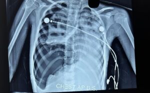

On laboratory investigations- There was leukocytosis,anemia,hypoalbuminemia,raised LDH,raised CRP-123mg/dl
Chest X-Ray showed right sided pneumothorax with multiple cavities.
Chest CT showed Pyonneumothorax with thin walled cavity in right lung.
DISCUSSION:
Necrotising pneumonia is a rare complication of pneumonia in children.
It is being recognised now a days due to increased use of CT scan.
It occurs in previusly healthy children due to high virulence of infecting organism.
Interaction between bacteria and viruses also play a role. The most common responsible organisma are Pneumococcus and Staphylococcus. It may be caused by Mycoplasma and Mycobacterium Tuberculosis.
PVL-Panton valentine leucocidin, secreted by MRSA is responsible in some cases. In such cases leucopenia rather than leucocytosis occurs with fast detrioration in clinical condition.
After infectin,vasculitis occurs in pulmonary vessels due to release of cytokines in which interleukin 8 driven neutrophils at inflammatory site,play an important role,leading to thrombosis,occlusion of vessels,activation of coagulation pathway, which causes liquefaction and necrosis, destruction of lung parenchyma takes place, and there occurs formation of multiple thin walled cysts. Somtimes gangrene occurs in whole lobe of lung.If the necrosis is near pleura,repture of pleura happens and pneumothorax develops. Pus then gets collected into pleural cavity and pyopneumothorax develops. This is the stage of Bronchopleural fistula. Bronchopleural fistula is usually a complication of chest tube drainage or surgical intervention but rarely occurs as an extension of disease itself.
TREATMENT:
It is mainly conservative with intravenous antibiotics with supportive care.Mechanical ventilation is required in case of respiratory failure. Chest tube insertion is avoided initially but situation of tension pneumothorax compells for chest tube drainage sometimes. .Fever persists for a long time. The duration of antibiotics may vary from 2- 6 weeks. I.V antibiotics should be given till the child is afebrile for at least 24 hours, imflammatory markers are coming down, hemodynamics are stable an oral feeds are tolerated.After that oral antibiotics may be needed for further 2- 3 weeks. Some children may require surgical intervention(VATS) to evacuate pus,sealing of Bronchopleural Fistula with FIBRIN GLUE or muscle flap. In some circumstances segmental resection of lung,lobectomy and even pneumenectomy may be required to save the child.
CONCLUSION:
Necrotising pneumonia is a very serious condtion ,requires intensive care ,sometimes with mechanical ventilation,chest tube drainage with fibrinolytics but mortality is very low with adequate treatment. Some cases may require prolonged antibiotics course with average duration of hospital of 28 days. In rare circumstances Video assistaed Thoracoscopic surgery is required.
Keywords:Necrotising pneumonia(NP),Pyopneumothorax,Tension pneumothorax,Bronchopleural Fistula(BPF),Thin walled lung cavity
REFERENCES:
1.Ramgopal S, Ivan Y, Medsinge A, Saladino RA. Pediatric necrotizing pneumonia and review of the literature. Pediatr Emerg Care. 2017;33:112–115. [PubMed] [Google Scholar]
2.McCarthy VP, Patamasucon P, Gaines T, Lucas MA. Necrotizing pneumonia in childhood. Pediatr Pulmonol. 1999;28:217–221. doi: 10.1002/(SICI)1099-0496(199909)28:3<217::AID-PPUL9>3.0.CO;2-R. [PubMed]
3.Gillet Y, Issartel B, Vanhems P, Fournet J-C, Lina G, Bes M, et al. Association between Staphylococcus aureus strains carrying gene for Panton-valentine leucocidin and highly lethal necrotising pneumonia in young immunocompetent patients. Lancet. 2002;359:753–759. doi: 10.1016/S0140-6736(02)07877-7. [PubMed] [CrossRef] [Google Scholar]
4.Kalaskar AS, Heresi GP, Wanger A, Murphy JR, Wootton SH. Severe necrotizing pneumonia in children, Houston, Texas, USA. Emerg Infect Dis. 2009;15:1696–1698. doi: 10.3201/eid1510.090589. [PMC free article] [PubMed] [CrossRef] [Google Scholar]
Please click on the link https://www.youtube.com/watch?v=X_C7Opms_fY
Please click on the link below
World Asthma Day is commemorated on every first Tuesday of May to increase awareness of asthma in public.
Today,7th May 2024 ,is World Asthma Day
There is a false perception among public that Asthma is a disease of adults and it does not occur in children.
The fact is that ,approximately 10-30 % children worldwide are suffering from Asthma and there are many hidden cases of Asthma in children ,where parents do not seek treatment from a qualified doctor who can diagnose Asthma in their children
The other problem is that ,it is very hard to accept the diagnosis of Asthma in children by their parents even if a qualified doctor tries to convince the diagnosis.
Ultimately ,child has to suffer the undiagnosed disease for years .
Dr Deo kumar Jha, MD, Pediatrician and Pulmonlogist,Delhi and NCR,Formery at AIIMS New Delhi
Asthma is the most common chronic respiratory disease in children.
It is both underdiagnosed and overdiagnosed in children
The standard treatment for childhood asthma is inhalational corticosteroid(ICS) in different doses according to the severity of asthma.
If the asthma is not controlled, on highest permissible doses of inhalational steroid,Long acting beta agonist(LABA) is added to control it, provided the technique of inhalation is correct,comorbidities have been addressed properly and allegen avoidence has been taken care of and adherence to treatment is good.
If it is not controlled on ICS+LABA,other add on options are LTRA(Monteleukast) and Tiotropium
If still the asthma is not controlled ,biologicals in the form of Omalizumab(IgE antagonist) and Meplozumab(IL5 antagonist) are given to control the asthma
Biologicals are costly with the disadvantages of adverse events and it is not widely available.
Asthma control is usually assessed by Asthma control test(ACT) ,Childhood asthma control test(CACT) and more easily by GINA guideline for control of asthma
Higher the ACT,CACT scores ,better is the control of asthma.
Researchers from the division of Pulmonology,department of Pediatrics,All India Institute of Medical Sciences,conducted an open label randomized control trial for a drug Azithromycin.Azithromycin is recommended drug by Global Initiative of Asthma(GINA) and British Thoracic Society(BTS) guideline for control of Asthma in adults.It improves spirometer parameter and reduces number of exacerbation of asthma in adults.There is no sufficient data for its use in children.
This the reason, researchers from Pediatric Pulmonology, division of the department of Pediatrcs AIIMS New Delhi, studied on 120 children between the age group of 5-15 years,mostly male(74% ) with poorly controlled asthma according to ACT and CACT.They divided these children into two groups.One group (n60) received Azithromycin in the dose of 10 mg/kg thrice weekly for 12 weeks along with standard treatment.The other group(n60) received only standard treatment.
The primary outcome was level of control of Asthma, according to ACT and CACT.Secondary outcomes were spirometry parameter,number of exacerbations,,Fractional excretion of NO(FeNO),throat swab culture positivity and adverse events
At the end of study period,the group who received Azithromycin along with standard care were having high ACT and CACT score (21.71 vs. 18.33; P < .001))indicating better asthma control.They also required less number of emergency visits due to asthma exacerbation and less use of oral or injectable steroids(0 vs. 1; P < .001).) ,higher number of good control of asthma by GINA guideline(41 vs. 10; P < .001).)
Spirometry parameters,throat swab culture ,FeNO reports and adverse events were not much different between two groups.
The benefits of Azithromycin was not different whether the child was suffering from eosinophilic or non eosinophilic asthma.
The study was published in CHEST.
CONCLUSION and BOTTOM LINE: Azithromycin in the dose of 10mg/kg,thrice weekly for 3 months may be added in treatment for children who could not achieve good control of asthma with standard therapy
REFERENCES:: Ghimire JJ, et al. Chest. 2022;doi:10.1016/j.chest.2022.02.025.
The most common chronic respiratory disease from which children all over the world suffer is ASTHMA.
Please click to know more for the better care of asthmatic children.
Please click below to watch DR.D.K.JHA discussing management of asthma in children in accordance with GINA guideline.
Please click below to watch DR.D.K JHA,talking invasive Pediatric Mechanical ventilation in a very simple and enjoying way