Injection to prevent HIV/AIDS Approved by USFDA,Dr Dev Kumar Jha,Pediatrician,Pediatric Pulmonologist and Intensivist Delhi and NCR

Injection to prevent HIV/AIDS Approved by USFDA,Dr Dev Kumar Jha,Pediatrician,Pediatric Pulmonologist and Intensivist Delhi and NCR

Dr D K JHA,Alumnus of AIIMS New Delhi, Child Specialist and Child Chest Super Specialist,L-27,L-Block,Lajpat Nagar,Sahibabad,Ghaziabad

Founder Director-Vitalis Super Speciality Children Hospital,D 64/1-Dilshad Colony ,Delhi 11095

Founder Director-Respiratory Foundation of India

Blood Donation and Blood Transfusion in Children-Dr Dev Kumar Jha,Pediatrician,Pediatric Pulmonologist & Intensivist Delhi ,Ghaziabad,NCR

DR.D.K.JHA,Alumnus of AIIMS,New Delhi, Child Specialist and Child Chest Super Specialist,L27,Near Kaali Mandir,L-Block,Lajpat Nagar Sahibabad, Ghaziabad

Founder Director -Vitalis Super Speciality Children Hospital,D 64/1-Dilshad Colony,Delhi 110095

Founder Director-Respiratory Foundation of India

Heat Wave and its Effect on us-Dr.D.K.JHA,Child Specialist & Child Chest Super Specialist,L-27,Near Kaali Mandir,Lajpat Nagar,Sahibabad,Delhi NCR,Phone-01203158081, 9971720128

Dr Dev Kumar Jha,Alumnus of AIIMS,New Delhi

Pediatric Pulmonologist & Intensivist ,Delhi & NCR

Founder Director-Dr. Dev CHILD HEALTH CARE & CHILD CHEST SUPER SPECIALITY CLINIC,L-27,

Near Kaali Mandir,Lajpat Nagar ,Sahibabad,Delhi NCR

Phone-010203158081,9971720128

website-www.childandchestdoctor.com

Founder Director of Respiratory Foundation of India

Founder Director of Vitalis Super Speciality Children Hospital ,D64/1-Dilshad Colony,New Delhi 110095

Lizard found in partly consumed food-what to do-Dr D K Jha,Child Specialist,Pediatric Pulmonologist & Intensivist,Rajendra Nagar ,Ghaziabad,Delhi NCR

Lizard poisoning

Collapse of Lung,Dr Dev Kumar Jha,M.D. Child specialist and child chest Superspecialist,Delhi and NCR

This child had been admitted in critical condtion in our Pediatric ICU.

The child was in respiratory distress with decompensated shock.

After initial fluid bolus,vasopressors were started.

The child  was maintaining saturation of 96% on room air but at the cost of increased respiratory rate and chest retraction.

The child was intubated and put on mechanical ventilator, to save the respiratory muscles from becoming fatigued.

The child became comfortable with no chest retraction, with saturation of 98% on ventilator.

After  few hours,saturation  decreased to 70% and ventilator settings were escalated to maintain the saturation.

On chest X-Xray examination, there were complete collapse of left lung which was detected to be responsible for decrease in saturation.

Chest phsiotherapy was started and the repeat chest X-Ray after few hours was normal.

The cause of lung collapse was mucus plug ,obstructiing the left main bronchus which was cleared by chest physiotherapy.

KEY MESSAGE– When the child is on ventilator and saturation falls, percuss and auscultate the chest.

If percussion note is dull and breath sound is diminished on that side ,suspect collapse of lung and do chest X-Ray to detect it.

Apart from mucus plug ,if the Endotracheal tube is placed inadvertantly low so as to enter the right main bronchus, there will be collapse of the left lung and hyperinflation of the right lung.

The tip of the endotracheal tube should lie 1.5 cm above the carina in case of children, which correspond between T2 to T4 vertebrae. It should be checked by chest X-Ray . Neck should be in neutral position,neither extended nor flexed whild doing the chest X-Ray.

On the other hand,if percussion note is hyper-resonant and breath sound is diminished,suspect Pneumothorax.

DR.DEV KUMAR JHA,MBBS(GOLD MEDALIST),M.D.(PEDIATRICS),FELLOWSHIP IN PEDIATRIC PULMONOLOGY,AIIMS,NEW DELHI


Necrotising Pneumonia with Bronchopleural Fistula,Case report-Dr Deo Kumar Jha,M.D., Child and Child Chest Specialist Ghaziabad,Delhi NCR

ABSTRACT;

Necrotising pneumonia is a serious and uncommon complication of pneumonia in children.It usually occurs in previously healthy children.It should be suspected when a child with pneumonia worsens clinically after 72 hours of appropriate  antibiotics. It may be complicated by pyopneumothorax and Broncopleural Fistula. It is diagnosed by radioimaging of chest and CECT Chest is the best modality.It needs long term antibiotics with long hospital stay.

CASE SUMMARY:

8 years male child came in our emergency with history of fever for 4 days with weakness and breathing difficulty for 2 days

On reaching hospital ,in the emergency department the child lost his consciousness

On examination ,the child was in shock and breath sound was diminished on right side of chest

The child had to put on ventilator immediately and then chest X-Ray was done which revealed right sided pneumothorax

Chest tube drainage was done immediately,which relieved pneumothorax

Appropriate antibiotics were started  but the condition of the child did not improve

Pus started coming out in the chest tube

CECT Chest was done which revealed cavitary lesions (thin walled cavities) with distortion of lung architecture with pyopneumothorax

There were appearance of air bubble in the chest tube which was not earlier after relieving of pneumothorax

It was then confirmed that there has been formation of Bronchopleural Fistula(BPF)

In view of  necrotising Pneumonia and BPF, antibiotics was upgraded to clindamycin and Meropenem

Ventilator settings was adjusted with low Ti. Low PEEP  and minimum Tidal Volume,


en

General condition improved gradually and child became afebrile,OG tube feeding was started while on ventilator

Condition became permissible to  extubate the child  after 2 weeks of  mechanical ventilation

While on ventilator ,the child had persistent metabolic alkalosis after the first report on ABG of respiratory acidosis before ventilation,there was persistent hypokalemia ,then one episode of respiratory alkalosis,all were managed properly. Child required one PRBC transfusion while on ventilator.

Chest tube could be removed after 3 weeks as BPF healed on conservative management

Now the child is on oral antibiotics and doing well

On laboratory investigations- There was leukocytosis,anemia,hypoalbuminemia,raised LDH,raised CRP-123mg/dl

Chest X-Ray showed right sided pneumothorax with multiple cavities.

Chest CT showed Pyonneumothorax with thin walled cavity in right lung.

DISCUSSION:

Necrotising pneumonia is a rare complication of pneumonia in children.

It is being recognised now a days due to increased use of CT scan.

It occurs in previusly healthy children due to high virulence of infecting organism.

Interaction between bacteria and viruses also play a role. The most common responsible organisma are Pneumococcus and Staphylococcus. It may be caused by Mycoplasma and Mycobacterium Tuberculosis.

PVL-Panton valentine leucocidin, secreted by MRSA is responsible in some cases. In such cases leucopenia rather than leucocytosis occurs with fast detrioration in clinical condition.

After infectin,vasculitis occurs in pulmonary vessels  due to release of cytokines in which interleukin 8 driven  neutrophils  at inflammatory site,play an important role,leading to thrombosis,occlusion of vessels,activation of coagulation pathway, which causes liquefaction and necrosis, destruction of lung parenchyma takes place, and there occurs formation of multiple thin walled cysts. Somtimes gangrene occurs in whole lobe of lung.If the necrosis is near pleura,repture of pleura happens and pneumothorax develops. Pus then gets collected into pleural cavity and pyopneumothorax develops. This is the stage of Bronchopleural fistula. Bronchopleural fistula is usually a complication of chest tube drainage or surgical intervention but rarely occurs as an extension of disease itself.

TREATMENT:

It is mainly conservative with intravenous antibiotics with supportive care.Mechanical ventilation is required in case of respiratory failure. Chest tube insertion is avoided initially but situation of tension pneumothorax compells for chest tube drainage sometimes. .Fever persists for a long time. The duration of antibiotics may vary from 2- 6 weeks. I.V antibiotics should be given till the child is afebrile for at least 24 hours, imflammatory markers are coming down, hemodynamics are stable an oral feeds are tolerated.After that oral antibiotics may be needed for further 2- 3 weeks. Some children may require surgical intervention(VATS) to evacuate pus,sealing of Bronchopleural Fistula with  FIBRIN GLUE or muscle flap. In some circumstances segmental resection of lung,lobectomy and even pneumenectomy may be required to save the child.

CONCLUSION:

Necrotising pneumonia is a very serious condtion ,requires intensive care ,sometimes with mechanical ventilation,chest tube drainage with fibrinolytics but mortality is very low with adequate treatment. Some cases may require prolonged antibiotics course with average duration of hospital of 28 days. In rare circumstances Video assistaed Thoracoscopic surgery is required.

Keywords:Necrotising pneumonia(NP),Pyopneumothorax,Tension pneumothorax,Bronchopleural Fistula(BPF),Thin walled lung cavity

REFERENCES:

1.Ramgopal S, Ivan Y, Medsinge A, Saladino RA. Pediatric necrotizing pneumonia and review of the literature. Pediatr Emerg Care. 2017;33:112–115. [PubMed] [Google Scholar]

2.McCarthy VP, Patamasucon P, Gaines T, Lucas MA. Necrotizing pneumonia in childhood. Pediatr Pulmonol. 1999;28:217–221. doi: 10.1002/(SICI)1099-0496(199909)28:3<217::AID-PPUL9>3.0.CO;2-R. [PubMed]

3.Gillet Y, Issartel B, Vanhems P, Fournet J-C, Lina G, Bes M, et al. Association between Staphylococcus aureus strains carrying gene for Panton-valentine leucocidin and highly lethal necrotising pneumonia in young immunocompetent patients. Lancet. 2002;359:753–759. doi: 10.1016/S0140-6736(02)07877-7. [PubMed] [CrossRef] [Google Scholar]

4.Kalaskar AS, Heresi GP, Wanger A, Murphy JR, Wootton SH. Severe necrotizing pneumonia in children, Houston, Texas, USA. Emerg Infect Dis. 2009;15:1696–1698. doi: 10.3201/eid1510.090589. [PMC free article] [PubMed] [CrossRef] [Google Scholar]

RECOGNITION OF SHOCK AT AN EARLY SATGE and Management ,DR.D.K.JHA,M.D.,Pediatrician and Pediatric Pulmonologist,Delhi

Assessment of hemodynamic conditions is most important in the management of critically ill patients

It is most important to pick up the condition of compensated shock and start treatment

Pediatric patients go into the stage of decompensated shock a bit late in comparasion to adult patients

Patients are managable in condition of decompesated shock if timely intervention is done.

Once the patient passes into irreversible shock it is very very difficult to revive and mortality is very high

How to recognise a patient clinically in a state of shock

STATE OF STABLE HEMODYNAMICS

Patient has clear consciousness

Periphery is warm and pink

Capillary refill time is 2 seconds or less

Pulse volume is good on palpation

Blood pressure is normal( between 5th to 95th centile for the age

Respiratory rate in the normal range for the age

Heart rate in the normal range for the age

Urine output normal for the age,1ml /kg/hour

STATE OF COMPENSATED SHOCK

Conscioussness inact

Periphery cool

Peripheral pulse ,low volume or thready

Capillary refill time more than 2 seconds

Blood pressure ,systolic is normal but diastolic is in rising trend,postural hypotension,narrow pulse pressure

Heart rate increased for the age

Respiratory rate increased for the age

Urine output reduced

STATE OF DECOMPENSATED SHOCK

Conscioussness-Restlessness or the patient is combative

Periphery on touch is cold and clammy

Capillary refill time is very prolonged like 5 seconds or more with or without mottling of skin

Peripheral pulse is very weak or may not be palpable at all even with great effort

Blood pressure -Hypotension,pulse pressure is 20 mm or less.Blood pressure may not be recordable

Heart rate-increased and in late stage decreased

Respiratory rate-Hyperpnea or Kussmaul breathing pattern(deep and sighy)

Urine output-oliguria or anuria

NORMAL RANGE of Respiratory rate

Premie-40-70/minute

0-3 months-30-60/minute

3-6 months-30-45/minute

6-12 months-25-40/minute

1-3 years-20-30/minute

3-6 years-20-25/minute

6-12 years-14-22/minute

>12 years 12-18/minute

NORMAL RANGE of heart rate- per minute

Premie 120-170

0-3 months 110-160

3-6 months 100-150

6-12 months 90-130

1-3 years 80-125

3-6 years 70-115

6-12 years 60-100

>12 years 60-100

HYPOTENSION is called when systolic blood pressure is below

60mm of Hg in NEW BORN,

70 mm of Hg between the age of 1 month to 1 year

70 mmHg+age in years multiplied by 2 ,between the age of 1- 10 years

90 mm Hg above the age of 10 years

Hypotension is also called when mean arterial pressure (MAP)is below 40+age in years multiplied by 1.5

MANAGEMENT:Management should start at the earliest, at the stage of compensated shock

First attention should be on airway and breathing and oxygen should be given if required to keep SPo2 95% and above

Life saving -for the circulation to be maintained, is fluid therapy

20 ml/kg of N/S or R/L shuold be given over 5-15 minutes and it should be pushed.It can be repeated twice if hydration,circulation and perfusion is not adequate.

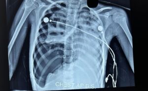
In the settings of obvious fluid loss like diarrhoea ,vomiting or hemorrhage ,repeated fluid administration should be done till the signs of fluid overload develop, in the form of tachycardia,bilateral deep inspiratory crackles over subscapular region,liver enlargement,engorgement of jugular vein or signs of pulmonary edema on chest X-Ray

R/L shuold not be used in case of a history of repeated vomiting

IV bolus should be repeated ,only when there is sign of improvement clinically and no sign of fluid overload.

Aggressive fluid therapy may be harmful and should not be given in certain situations like shock in the settings of severe acute malnutrition,severe anemia,compensated shock with high fever with no dehydration or obvious fluid loss(Dengue fever),cardiogenic shock(ductal dependent congenital heart disease in newborn),obstructive shock(tension pneumothorax,cardiac temponade,)

SIGHNS OF CARDIOGENIC SHOCK-Tachycardia,engorged jugular vein,bilateral deep inspiratory crackles over subscapular regions,gallop rhythm,liver enlargement,signs of pulmonary edema on chest X-Ray.

In these cicumstances,crystalloids(N/S or R/L) should be given in the dose of 5-10 ml per kg over 15-30 minutes once then switch over to vasopressors

In case of poor response or no response to fluid therapy,swith over to vasopressures without delay.

If the periphery is cold,give DOPAMINE/EPINEPHRINE

If the perphery is warm give NORADRENALINE

In case of myocardial dysfuntion with maintained blood pressure,give DOBUTAMINE

In case of myocardial dysfuntion with increased peripheral resistance,use MILRINONE

Easy preparation and administration of vasopressures

Dopamine/Dobutamine 6 mg/kg-dilute in 100 ml of D5-1 ml/hour of this will deliver 1 mcg/kg/minute=Dose is 5-20 mcg/kg/minute

EPINEPHRINE0.6mg/kg of body weight,dilute in 100 ml of D5-1ml/hour will deliver 0.1mcg/kg/minute=Dose 0.05 to 0.2mcg/kg/minute,in severe cases upto 1mcg/kg/minute

Norepinephrine 0.6 mg/kg,dilute in 100 ml D5,1ml/hour will deliver 0.1mcg/kg/minute=Dose 0.1-1mcg/kg/hour

REFERENCES1.;Harriet Lane 21 edition

2. CDC guideline on management of shock in children

3.Uptodate-management of shock in children,2021
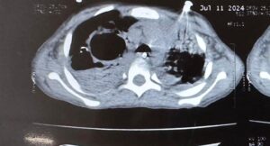
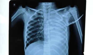
Dengue fever/MIS-C,DR D.K.JHA,M.D.,Pediatrician and Pediatric Pulmonologist Delhi

In the period of october 2021,both Dengue fever and Multi system inflammatory syndrome in children(MIS-C) are being seen in children in Delhi,India

Clinical and laboratory features are overlapping for both these diseases

It is important to differentiate between these as management entirely differs for both

In case of Dengue fever the cornerstone of management is aggressive fluid therapy with crystalloid and colloid with ionotrops if fluid therapy does not work and platelet transfusion if needed.

In cases of MIS-C, the cornerstone of management is steroids and IVIG(Immunoglobulins).Aggressive fluid management may be detrimental in cases of shock with cardiac dysfunction.

Fever are common in both but swellings of feet and hands,diarrhoea,conjuntival injections and altered sensorium along with the laboratory findings of hyperinflammation like highly raised CRP,Leukocytosis,raised D-Dimer are pointers towards MIS-C .In this situations,anti COVID antibody should be done and if positive ,confirms the diagnosis of MIS-C

If fever is associated with vomiting ,erythmatous rashes,myalgia along with the laboratory findings of leucopenia ,severe thrombocytopenia,hemoconcentration , raised serum ferritin level,it points towards the diagnosis of Dengue fever and NS1 antigen and or anti Den IgM should be done which when positive confirms the diagnosis of Dengue fever

In comparision to MIS-C,serum Ferritin level is higher in Dengue fever

References:

1. Ahmed M, Advani S, Moreira A, et al. Multisystem
inflammatory syndrome in children: A systematic review. E
Clin Med. 2020;26:100527.

2.Mishra S, Ramanathan R, Agarwalla SK. Clinical profile of
dengue fever in children: A study from southern Odisha,
India. Scientifica (Cairo). 2016;2016:6391594

3.Indian Pediatrics,volume 58,15 October,2021

INVASIVE PEDIATRIC MECHANICAL VENTILATION MADE SIMPLE BY DR.D.K.JHA,M.D.,Pediatric Pulmonologist and Respiratory Intensivist-Delhi

Please click below to watch DR.D.K JHA,talking invasive Pediatric Mechanical ventilation in a very simple and enjoying way