Fellowship in Pediatric Pulmonology & Bronchoscopy
By Respiratory Foundation of India
Whatsapp for registration -9899186848, 9873446805
For insight ,visit our website-www.rfoindia.com
Fellowship in Pediatric Pulmonology & Bronchoscopy
By Respiratory Foundation of India
Whatsapp for registration -9899186848, 9873446805
For insight ,visit our website-www.rfoindia.com
Dr D K JHA,Alumnus of AIIMS New Delhi, Child Specialist and Child Chest Super Specialist,L-27,L-Block,Lajpat Nagar,Sahibabad,Ghaziabad
Founder Director-Vitalis Super Speciality Children Hospital,D 64/1-Dilshad Colony ,Delhi 11095
Founder Director-Respiratory Foundation of India
DR.D.K.JHA,Alumnus of AIIMS,New Delhi, Child Specialist and Child Chest Super Specialist,L27,Near Kaali Mandir,L-Block,Lajpat Nagar Sahibabad, Ghaziabad
Founder Director -Vitalis Super Speciality Children Hospital,D 64/1-Dilshad Colony,Delhi 110095
Founder Director-Respiratory Foundation of India
In a study at University of Copenhagen and Bispebjerg Hospital ,40 COPD patients and 20 control were included in Double blind placebo controlled randomised study.
2gm per day of vitamin B3(Nicotinamide riboside) were given to one group and placebo was given to other group
After 12 weeks inflammatory marker interleukin 8,IL8 was measured in both group
The group receiving VITAMIN B3 showed 53% reduction in the level of IL8 as compared to the group which did receive placebo
After another 12 weeks this reduction was 63%
NAD which reduces with age was higher in the group receiving VITAMIN B3 .
NAD reduction is correlated with aging process and DNA methylation.With aging the level of NAD reduces in body.
This study shows that treatment with VITAMIN B3 may be beneficial in diseases of lungs where inlmmations due to infections or pollutants is responsible for lung damages and disease manifestations
REFERENCES: Published online 8.30pm,27.11.24
Dr Dev Kumar Jha
Fellowship in Pediatric Pulmonology,AIIMS,New Delhi
M.B.B.S(Gold Medalist)
M.D.(Pediatrics)
Recipient of Bharat Ratna Dr APJ Abdul Kalam Azad National Icon Award 2021 inHealth Services
Vice President-Pediatric Respiratory Society Delhi
Visiting Consultant-Yashoda Super Speciality Hospital,Nehru Nagar,Kaushambi, Yashoda Medicity,Indirapuram,Ghaziabad Delhi NCR
Head of Pediatric Pulmonology Division, Department of Pediatrics,Santosh Medical College Hospital,Ghaziabad,Delhi NCR
Director & Senior Consultant- Dr Dev CHILD HEALTH CARE & CHILD CHEST SUPER SPECIALITY CLINIC,Lajpat Nagar,Sahibabad,Delhi-NCR, Contact -01203158081,9971720128,9873446805
Coordinator & Director- DDCD-Dr Dev Classes for Doctors,Whatsapp 9899186848
ABSTRACT:
Tropical Pulmonary eosinophilia is a rare disease affecting multiple organs but there is more involvement of lungs.It ia caused by hypersensitivity reactions to trapped microfilariae of Wuchereria bancrofti(W.bancrofti) and Brugia malayi(B.malayi) in the lungs. I report a case of TPE(Tropical Pulmonary eosinophilia in a child who presented similar to Pulmonary tuberculosis,diagnosed and treated timely with good response.
KEYWORDS Tropical pulmonary eosinophilia,hypersesitivity reactions, mocrofilaria,Filaria
INTRODUCTION:
Tropical Pulmonary eosinophilia as a disease entity was first described in 1940 and the term Pseudotuberculosis with eosinophilia was given.In 1943 Weingarten coined the term TPE(Tropical Pulmonary eosinophilia) for this disease. The disease occurs in costal India like Maharashtra,Kerala,Goa and odisha.It has also been found in some areas of Bihar( prevalence of 9% in jail inmates of Patna).A prevalence of 0.5% among children of Tamil Nadu has been found.It occurs in filaria endemic zones of South America,Africa and South East Asia.The common presentation is cough,difficulty in breathing,fever and weight loss. On investigation there is Leucocytosis with eosinophilia,raised serum IgE and raised specific serum IgE and IgG against Filaria antigen. Treatment is 3-4 weeks course of DEC(Diethylecarbamzepine)
CASE SUMMARY:
9 years old female child came to my OPD with complaint of fever off and on for 2-3 months,cough for 2 months,difficulty in breathing for 1 month ,breathless on exertion for 1 month.
There was no contact history for tuberculosis in the family or relatives
She was taking treatment from different doctors in past 3 months
She was also loosing her weight in last 2 months and she was easily fatigued in last 1 month
on clinical examination she was underweight and anemic,there was significant cervical lymphadenopathy ,no clubbing,no cyanosis and on chest auscultation there were bilateral wheezes and crackles.Abdominal examination showed hepatsplenomegaly
On investigation
Hb 14.3g/dl,
TLC-50,000/cmm,
Neutrophil 28%,lymphocytes10%,
Eosinophil 61%
Basophil 0%,ESR18mm/1H,
Peripheral smear examination-leucocytes increased in number with normal distribution, platelets adequate,no blast cells, malaria parasites amd microfilariae absent
Chest X-Ray-multiple bilateral miliary mottling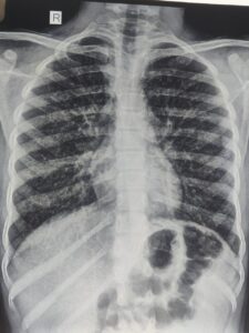
I thought it may be a case of
1.Tuberculosis with some parasitic infections,
2.Asthma with ABPA,
3.hypereosinophilic syndrome,
4.Tropical Pulmonary Eosinophilia
Then , I investigated further
GA for CBNAAT -Negative,Montoux test -negative
Serum IgE -3389U/L,Serum specific filaria antibody IgE and Ig G were raised
Then,I Put the child on DEC 5 mg/kg/day for 4 weeks orally and child started improving after 1 week of initiating DEC
Repeat Serum IgE after 4 weeks was normal ,TLC came down to normal range, and repeat chest X-Ray at that time was normal,child started gaining weight
DISCUSSION:
TPE is usually seen between the age group of 15-45 years .It is rare to see in children less than 15 years ,which was our case. The male:female is 4;1 which is against our case.The clinical features are malaise,fatigue,cough ,chest pain, breathlessness which are seen more during night.Clinical findings in chest may be wheezes and crackes but in 20% cases,there may be no findings in chest.It may involve any system apart from chest leading to the findings of cervical lymphadenopathy,hepatomegaly,splenomegaly,hepatosplenomegaly
The pathology involves hypersensitivity reactions type I,III,and IV to microfilariae.It is due to release of microfilariae from lymphatics which get entrapped into pulmonary circulation.Degranulation of eosinophils, release major basic proteins along with other granules which destruct microfilariae as well as respiratory and terminal bronchioles with lung parenchyma.In long standing untreated cases fibrosis develops.
Lung function test shows mixed obstructive and restrictive pattern of lung disease.
Chest radiograph shows raticuonodular opacities in mid zone and lower zone but as the disease progresses,there are miliary mottling all over the lung fields. Chest X-Ray may be normal in about 20% cases.CT chest may show mediastinal lymphadenopathy,consolidation,bronchiectasis,cavitation and Pleural effision.
CBC shows leococytosis with marked eosinophilia,AEC is usually more than 3000/cmm,Total serum IgE and specific IgE and IgG against Filaria is raised.
ELISA test detecting 0g4c3 antigen is sensitive and specific to W.bancrofti and Sanwich ELISA detecting recombinant antigen Bm-SXP-1 is specific to B.malayi
Trearment is DEC,5mg/kg/day for 4 weeks.Steroids may have a role but dose and duration is yet to be known
Our case is well fitting into the diagnostic criteria
CONCLUSION: TPE is a hypersensitive reaction to W.Bancrofti and B.malayi microfilariae mainly type I, causing eosinophilic alveolitis with inflammation of terminal an d respiratory bronchioles.Diagnosed by clinical features ,mainly night symptoms of cough and dyspnea,peripheral eosinophilia which has diurnal variation and paradoxically low level during night,raised serum IgE and specific IgE and IgG against filaria ,infiltrates on chest radiograph.Treatment is oral DEC for 4 weeks but there is 20% relapse within 5 years. The disease closely resembles Pulmonary tuberculosis and asthma.Untreated and partialy treated case may progress to Interstitial lung disease(ILD)
REFERENCES:
1.Indian J Med Res. 2013 Sep; 138(3): 295–302.
2. Udwadia FE. Herzog H, editor. Tropical eosinophilia. Pulmonary eosinophilia: progress in pulmonary research. 1975;VII:35–155. Basel: S Karger
3.Neva FA, Kaplan AP, Pacheco G, Gray L, Danaraj TJ. Tropical eosinophilia. A human model of parasitic immunopathology, with observations on serum IgE levels before and after treatment. J Allergy Clin Immunol. 1975;55:422–9
4. Frimodt-Moller C, Barton RM. A pseudo-tuberculosis condition associated with eosinophilia. Indian Med Gaz. 1940;75:607–13
 This child had been admitted in critical condtion in our Pediatric ICU.
The child was in respiratory distress with decompensated shock.
After initial fluid bolus,vasopressors were started.
The child was maintaining saturation of 96% on room air but at the cost of increased respiratory rate and chest retraction.
The child was intubated and put on mechanical ventilator, to save the respiratory muscles from becoming fatigued.
The child became comfortable with no chest retraction, with saturation of 98% on ventilator.
After few hours,saturation decreased to 70% and ventilator settings were escalated to maintain the saturation.
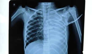
On chest X-Xray examination, there were complete collapse of left lung which was detected to be responsible for decrease in saturation.
Chest phsiotherapy was started and the repeat chest X-Ray after few hours was normal.
The cause of lung collapse was mucus plug ,obstructiing the left main bronchus which was cleared by chest physiotherapy.
KEY MESSAGE– When the child is on ventilator and saturation falls, percuss and auscultate the chest.
If percussion note is dull and breath sound is diminished on that side ,suspect collapse of lung and do chest X-Ray to detect it.
Apart from mucus plug ,if the Endotracheal tube is placed inadvertantly low so as to enter the right main bronchus, there will be collapse of the left lung and hyperinflation of the right lung.
The tip of the endotracheal tube should lie 1.5 cm above the carina in case of children, which correspond between T2 to T4 vertebrae. It should be checked by chest X-Ray . Neck should be in neutral position,neither extended nor flexed whild doing the chest X-Ray.
On the other hand,if percussion note is hyper-resonant and breath sound is diminished,suspect Pneumothorax.
DR.DEV KUMAR JHA,MBBS(GOLD MEDALIST),M.D.(PEDIATRICS),FELLOWSHIP IN PEDIATRIC PULMONOLOGY,AIIMS,NEW DELHI
ABSTRACT;
Necrotising pneumonia is a serious and uncommon complication of pneumonia in children.It usually occurs in previously healthy children.It should be suspected when a child with pneumonia worsens clinically after 72 hours of appropriate antibiotics. It may be complicated by pyopneumothorax and Broncopleural Fistula. It is diagnosed by radioimaging of chest and CECT Chest is the best modality.It needs long term antibiotics with long hospital stay.
CASE SUMMARY:
8 years male child came in our emergency with history of fever for 4 days with weakness and breathing difficulty for 2 days
On reaching hospital ,in the emergency department the child lost his consciousness
On examination ,the child was in shock and breath sound was diminished on right side of chest
The child had to put on ventilator immediately and then chest X-Ray was done which revealed right sided pneumothorax
Chest tube drainage was done immediately,which relieved pneumothorax
Appropriate antibiotics were started but the condition of the child did not improve
Pus started coming out in the chest tube
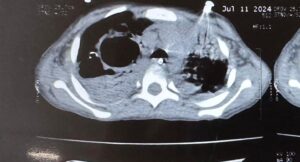
CECT Chest was done which revealed cavitary lesions (thin walled cavities) with distortion of lung architecture with pyopneumothorax
There were appearance of air bubble in the chest tube which was not earlier after relieving of pneumothorax
It was then confirmed that there has been formation of Bronchopleural Fistula(BPF)
In view of necrotising Pneumonia and BPF, antibiotics was upgraded to clindamycin and Meropenem
Ventilator settings was adjusted with low Ti. Low PEEP and minimum Tidal Volume,

 en
en
General condition improved gradually and child became afebrile,OG tube feeding was started while on ventilator
Condition became permissible to extubate the child after 2 weeks of mechanical ventilation
While on ventilator ,the child had persistent metabolic alkalosis after the first report on ABG of respiratory acidosis before ventilation,there was persistent hypokalemia ,then one episode of respiratory alkalosis,all were managed properly. Child required one PRBC transfusion while on ventilator.
Chest tube could be removed after 3 weeks as BPF healed on conservative management
Now the child is on oral antibiotics and doing well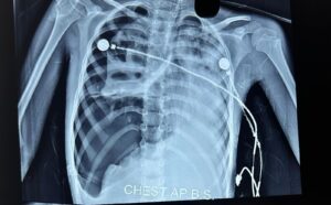

On laboratory investigations- There was leukocytosis,anemia,hypoalbuminemia,raised LDH,raised CRP-123mg/dl
Chest X-Ray showed right sided pneumothorax with multiple cavities.
Chest CT showed Pyonneumothorax with thin walled cavity in right lung.
DISCUSSION:
Necrotising pneumonia is a rare complication of pneumonia in children.
It is being recognised now a days due to increased use of CT scan.
It occurs in previusly healthy children due to high virulence of infecting organism.
Interaction between bacteria and viruses also play a role. The most common responsible organisma are Pneumococcus and Staphylococcus. It may be caused by Mycoplasma and Mycobacterium Tuberculosis.
PVL-Panton valentine leucocidin, secreted by MRSA is responsible in some cases. In such cases leucopenia rather than leucocytosis occurs with fast detrioration in clinical condition.
After infectin,vasculitis occurs in pulmonary vessels due to release of cytokines in which interleukin 8 driven neutrophils at inflammatory site,play an important role,leading to thrombosis,occlusion of vessels,activation of coagulation pathway, which causes liquefaction and necrosis, destruction of lung parenchyma takes place, and there occurs formation of multiple thin walled cysts. Somtimes gangrene occurs in whole lobe of lung.If the necrosis is near pleura,repture of pleura happens and pneumothorax develops. Pus then gets collected into pleural cavity and pyopneumothorax develops. This is the stage of Bronchopleural fistula. Bronchopleural fistula is usually a complication of chest tube drainage or surgical intervention but rarely occurs as an extension of disease itself.
TREATMENT:
It is mainly conservative with intravenous antibiotics with supportive care.Mechanical ventilation is required in case of respiratory failure. Chest tube insertion is avoided initially but situation of tension pneumothorax compells for chest tube drainage sometimes. .Fever persists for a long time. The duration of antibiotics may vary from 2- 6 weeks. I.V antibiotics should be given till the child is afebrile for at least 24 hours, imflammatory markers are coming down, hemodynamics are stable an oral feeds are tolerated.After that oral antibiotics may be needed for further 2- 3 weeks. Some children may require surgical intervention(VATS) to evacuate pus,sealing of Bronchopleural Fistula with FIBRIN GLUE or muscle flap. In some circumstances segmental resection of lung,lobectomy and even pneumenectomy may be required to save the child.
CONCLUSION:
Necrotising pneumonia is a very serious condtion ,requires intensive care ,sometimes with mechanical ventilation,chest tube drainage with fibrinolytics but mortality is very low with adequate treatment. Some cases may require prolonged antibiotics course with average duration of hospital of 28 days. In rare circumstances Video assistaed Thoracoscopic surgery is required.
Keywords:Necrotising pneumonia(NP),Pyopneumothorax,Tension pneumothorax,Bronchopleural Fistula(BPF),Thin walled lung cavity
REFERENCES:
1.Ramgopal S, Ivan Y, Medsinge A, Saladino RA. Pediatric necrotizing pneumonia and review of the literature. Pediatr Emerg Care. 2017;33:112–115. [PubMed] [Google Scholar]
2.McCarthy VP, Patamasucon P, Gaines T, Lucas MA. Necrotizing pneumonia in childhood. Pediatr Pulmonol. 1999;28:217–221. doi: 10.1002/(SICI)1099-0496(199909)28:3<217::AID-PPUL9>3.0.CO;2-R. [PubMed]
3.Gillet Y, Issartel B, Vanhems P, Fournet J-C, Lina G, Bes M, et al. Association between Staphylococcus aureus strains carrying gene for Panton-valentine leucocidin and highly lethal necrotising pneumonia in young immunocompetent patients. Lancet. 2002;359:753–759. doi: 10.1016/S0140-6736(02)07877-7. [PubMed] [CrossRef] [Google Scholar]
4.Kalaskar AS, Heresi GP, Wanger A, Murphy JR, Wootton SH. Severe necrotizing pneumonia in children, Houston, Texas, USA. Emerg Infect Dis. 2009;15:1696–1698. doi: 10.3201/eid1510.090589. [PMC free article] [PubMed] [CrossRef] [Google Scholar]
 Case summary
11 years female child came to our OPD with complaints of 4 episodes of sudden loss of consciousness,what the parents called,fainting attacks 10 months back.
On all occasions,there was no abnormal body movement and each episode lasted for less than a minute.
The child was completely normal after the episodes.
In the last 3 months ,the parents noticed easy fatiguebility in the child.
The child was experiencing breathlessness on little exertion.
ON clinical examination
There was no clubbing,respiratory rate and pulse rate were normal,SPo2 on room air was 98%
On chest auscultation,breath sounds were normal and there was no adventitious sound.
ON auscultaion of heart-P2 was loud
INVESTIGATION;Chest X-Ray was normal,Echocardiography revealed enlarged right atrium and right ventricle and bulging of ineratrial septum towards left atrium.
Pulmonary artery pressure was 60 mm of Hg.
CT Pulmonary angiography revealed filling defects in pulmonary arteries supplying to the right upper lobe of the lung at multiple sites and few sites on the left lung also.
Pulmonary trunk was dilated and heart chambers of right side were enlarged.
On further investigations-Factor V Leiden,Protein C,Protein S were negative but ANA was positive(1:320).
Anti ds antibody,anti smith antibody and ENA(Extractable nuclear antigen) were negative.
Lupus anti coagulant elevated,anti phospholipid antibodies in the form of anticardiolipin antibody and anti beta 2 glycoprotein 1 antibodies on two occasions ,3 months apart were positive.
DIAGNOSIS
According to EULAR 2006(,European Alliance of Association of Rheumatology) criteria the diagnosis was made of anti phospholipid syndrome–APS
TREATMENT:
Enoxaperin 1 mg /kg/dose 12 hourly was started for pulmonary thrombosis along with Sildenafil 12.5 mg three times daily for pulmonary hypertension.
Discussion:
we present a case of a female child with pulmonary hypertension due to pulmonary thrombosis with antiphospholipid syndrome.
It is important to recognise the association of Pulmonary thrombosis with Antiphopholipid syndrome(APS)
Pulmonary hypertension is dianosed if Pulmonary artery pressure is more than 25 mm of Hg IN CHILDREN > 3 months.
Symptoms include fatigue,exertional dyspnea and syncope.
Treatment includes PED5 inhibitor like sildenafil
It is not uncommon in adults but rare in children with frequency of less than 1 per million children.
APS is diagnosed when at least one clinical condition like thrombosis or morbidities of pregnancy and at least one laboratory finding (Anti lupus anticoagulant ,Antiphospholipid antibody) are positive on 2 separate occasions 12 weeks apart.It is more common in female and treatment is anticoagulants for thrombosis.
In children mostly it is secondary to autoimmune diseases and 20% primary APS develop lupus.
CONCLUSION: Since the association of APS with PH is rare ,it will be beneficial to report such case as early recognition and treatment may improve the outcome
REFERENCES:
bman SH, Hansmann G, Archer SL, et al.: Pediatric pulmonary hypertension: guidelines from the American Heart Association and American Thoracic Society. Circulation. 2015, 132:2037-99. 10.1161/CIR.0000000000000329
Hansmann G: Pulmonary hypertension in infants, children, and young adults. J Am Coll Cardiol. 2017, 69:2551-69. 10.1016/j.jacc.2017.03.575
Fraisse A, Jais X, Schleich JM, et al.: Characteristics and prospective 2-year follow-up of children with pulmonary arterial hypertension in France. Arch Cardiovasc Dis. 2010, 103:66-74. 10.1016/j.acvd.2009.12.001
Cerro MJ, Abman S, Diaz G, et al.: A consensus approach to the classification of pediatric pulmonary hypertensive vascular disease: report from the PVRI Pediatric Taskforce, Panama 2011. Pulm Circ. 2011, 1:286-98. 10.4103/2045-8932.83456
Berger RM, Beghetti M, Humpl T, et al.: Clinical features of paediatric pulmonary hypertension: a registry study. Lancet. 2012, 379:537-46. 10.1016/S0140-6736(11)61621-8
Barst RJ, McGoon MD, Elliott CG, Foreman AJ, Miller DP, Ivy DD: Survival in childhood pulmonary arterial hypertension: insights from the registry to evaluate early and long-term pulmonary arterial hypertension disease management. Circulation. 2012, 125:113-22. 10.1161/CIRCULATIONAHA.111.026591
Avitabile CM, Vorhies EE, Ivy DD: Drug treatment of pulmonary hypertension in children. Paediatr Drugs. 2020, 22:123-47. 10.1007/s40272-019-00374-2
Rosina S, Chighizola CB, Ravelli A, Cimaz R: Pediatric antiphospholipid syndrome: from pathogenesis to clinical management. Curr Rheumatol Rep. 2021, 23:10.1007/s11926-020-00976-7
Madison JA, Zuo Y, Knight JS: Pediatric antiphospholipid syndrome. Eur J Rheumatol. 2020, 7:3-12. 10.5152/eurjrheum.2019.19160
Tektonidou MG, Andreoli L, Limper M, et al.: EULAR recommendations for the management of antiphospholipid syndrome in adults. Ann Rheum Dis. 2019, 78:1296-304. 10.1136/annrheumdis-2019-215213