CAUSES OF HAPPY HYPOXIA IN COVID 19,Dr.Dev,M.D.,Pediatrician and Pediatric Pulmonologist,Sahibabad,Ghaziabad,Delhi NCR

CAUSES OF HAPPY HYPOXIA IN COVID 19,Dr.Dev,M.D.,Pediatrician and Pediatric Pulmonologist,Sahibabad,Ghaziabad,Delhi NCR

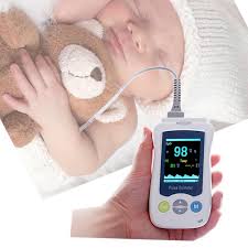
Many patients suffering from COVID 19 are hypoxemic but not in distress and breathing comfortably in room air.This stage is called happy hypoxia.Hpoxemia is detected only after high index of suspician leads to SpO2 monitoring.

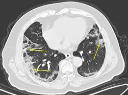
Whenever there is less ventilation into the lung,there occurs a  vasoconstriction in the pulmonary vessels as a compensatory response in poorly ventilated area of the lung..This is called hypoxemic Pulmonary vasocontrictor response(HPV).It has been seen that in COVID19 ,this HPV response is diminished with formation of intrapulmonary shunt(right to left) as demontrated by dual energy CT scan. This vasoplegic response is triggered by vasodilator prostaglandin,bradykinin and cytokines.

SARS Cov 2 virus,responsible for COVID 19, damages the endothelium of lung which express angiotensin converting enzyme II receptors, leading to formation of microthrombi in lung capillaries.There is extensive thrombi formation in arteries and capillaries leading to blockage of capillaries and arteries causing ventilation/perfusion mismatch and hypoxemia.This stage is called AVDS(acute vascular distress syndrome)

SARS Cov2 virus damages type 2 pneumocytes and in effect, basement membrane is denued which becomes covered by hyaline membrane consisting of fibrin and dead cells which impaires gas exchange.As the diffusion barrier becomes more pronounced,blood flow through pulmonary vessels increase during exertion.So there is less time for gas exchange during exertion causing hypoxemia.

In normal situations whenever there is less oxygen in the blood,it is sensed by carotid bodies which is situated at the bifurcation of commonl carotid artery..It gives signal to the respiratory centre situated in medulla oblongata which in turn sends signal to diaphragm via phrenic nerve to increase ventilation which is perceived as duspnea by patients.When the PaO2 is between 90 mmHg to 60 mmHg,there is no  such response but as soon as the PaO2 drops below 60 mmHg ,immediately there is a signal exponentially, to increase the ventilation.But for this ventilatory response to occur, when the level of PaO2 is between 40mmHg to 60 mmHg,PaCo2 level must increase to above 39 mmHg to stimulate the carotid bodies.

 SARS COV2 VIRUS BLOCKS the sensitivity of angiotensin converting enzyme 2 receptor present in carotid body .Therefore it can not sense hypoxia or hypercarbia unless it becomes critical.So,there is no signal to increase the ventilation and patients remain comfortable till the hypoxia becomes critical.This stage is called happy hypoxia.

In the early stage of the disease,lung parenchyma is minimally involved which is evident by ground glass opacity in the peripheral regions of  lung.In this stage ,vital capacity is maintained ,there is high compliance and low elastance of lung called L-Phenotype.At this stage there is low  ventilatory response as the hypoxia is vascular in origin due to impairement of HPV  and there is no dyspnea.

As the disease progresses,there is extensive involvement of lung parenchyma with increased ventilatory response,increased permeability of pulmonary capillaries with accumulation of fluid in interstitium(interstitial edema)and inspiratory pressure becomes more negative.This stage is called H-Phenotype with high elastance and low compliance.The transition from L-phenotype to H-Phenotype is precipitaed  by more negative inspiratory pressure created by forceful inspiratory effort by patient during spontaneous breathing,called patient self inflected lung injury(P-SILI)

In case of fever, the oxygen dissociation curve is shifted towards right side, causing low SaO2 for a given PaO2, making the sensing of SpO2 by pulse oxymetry inaccurate as ,at low PaO2 level, SpO2 can underread by about 7 % if SaO2 is below 80%

In case of Diabetes and age more than 65 years the sensitivity of carotid body becomes less causing less ventilatory response.


IN NUTSHELL, at the initial stage of disease,there is minimal involvement of lung parenchyma,primarily the disease affects pulmonary vessels with less  HPV response,AVDS,with development of rifht to left intrapulmonary shunt causing hypoxia,the virus decreases the sensitivity of carotid body to sense hypoxia and increase ventilatory response,shifting of oxygen dissociation curve to right causing low SaO2 for a given PaO2 due to high fever, all leading to less ventilatory response and patients remains comfortable till the critical stage.


REFERENCES:

1. Guan W, Ni Z, Hu Y, et al. Clinical Characteristics of Coronavirus Disease 2019 in China. N Engl J Med. 2020;382(18):1708-1720. doi:10.1056/NEJMoa2002032

2. Mahjoub Y, Rodenstein DO, Jounieaux V. Severe Covid-19 disease: rather AVDS than ARDS? Crit Care. 2020;24(1):327. doi:10.1186/s13054-020-02972-w

3. Lang M, Som A, Mendoza DP, et al. Hypoxaemia related to COVID-19: vascular and perfusion abnormalities on dual-energy CT. Lancet Infect Dis. Published online April 30, 2020. doi:10.1016/S1473-3099(20)30367-4

4. Dhont S, Derom E, Van Braeckel E, Depuydt P, Lambrecht BN. The pathophysiology of ‘happy’ hypoxemia in COVID-19. Respir Res. 2020;21(1):198. doi:10.1186/s12931-020-01462-5

5. Ackermann M, Verleden SE, Kuehnel M, et al. Pulmonary Vascular Endothelialitis, Thrombosis, and Angiogenesis in Covid-19. N Engl J Med. 2020;383(2):120-128. doi:10.1056/NEJMoa2015432

6. Mahjoub Y, Rodenstein DO, Jounieaux V. Severe Covid-19 disease: rather AVDS than ARDS? Crit Care. 2020;24(1):327. doi:10.1186/s13054-020-02972-w

7. Mason RJ. Pathogenesis of COVID-19 from a cell biology perspective. Eur Respir J. 2020;55(4). doi:10.1183/13993003.00607-2020

8. Mo X, Jian W, Su Z, et al. Abnormal pulmonary function in COVID-19 patients at time of hospital discharge. Eur Respir J. 2020;55(6):2001217. doi:10.1183/13993003.01217-2020

9. Hopkins SR. Exercise Induced Arterial Hypoxemia: The role of Ventilation-Perfusion Inequality and Pulmonary Diffusion Limitation. In: Roach RC, Wagner PD, Hackett PH, eds. Hypoxia and Exercise. Advances in Experimental Medicine and Biology. Springer US; 2007:17-30. doi:10.1007/978-0-387-34817-9_3

10. Masi P, Bagate F, d’Humières T, et al. Is hypoxemia explained by intracardiac or intrapulmonary shunt in COVID-19-related acute respiratory distress syndrome? Ann Intensive Care. 2020;10(1):108. doi:10.1186/s13613-020-00726-z

11. Reynolds AS, Lee AG, Renz J, et al. Pulmonary Vascular Dilatation Detected by Automated Transcranial Doppler in COVID-19 Pneumonia. Am J Respir Crit Care Med. Published online August 6, 2020:rccm.202006-2219LE. doi:10.1164/rccm.202006-2219LE

12. Mohan R, Duffin J. The effect of hypoxia on the ventilatory response to carbon dioxide in man. Respir Physiol. 1997;108(2):101-115. doi:10.1016/S0034-5687(97)00024-8

13. Gattinoni L, Chiumello D, Caironi P, et al. COVID-19 pneumonia: different respiratory treatments for different phenotypes? Intensive Care Med. 2020;46(6):1099-1102. doi:10.1007/s00134-020-06033-2

14. Weisbrod CJ, Eastwood PR, O’Driscoll G, Green DJ. Abnormal ventilatory responses to hypoxia in Type 2 diabetes. Diabet Med J Br Diabet Assoc. 2005;22(5):563-568. doi:10.1111/j.1464-5491.2005.01458.x

15. Kronenberg RS, Drage CW. Attenuation of the ventilatory and heart rate responses to hypoxia and hypercapnia with aging in normal men. J Clin Invest. 1973;52(8):1812-1819. doi:10.1172/JCI107363

16. Tobin MJ, Laghi F, Jubran A. Why COVID-19 Silent Hypoxemia Is Baffling to Physicians. Am J Respir Crit Care Med. 2020;202(3):356-360. doi:10.1164/rccm.202006-2157CP

17. Kelman GR. Digital computer subroutine for the conversion of oxygen tension into saturation. J Appl Physiol. 1966;21(4):1375-1376. doi:10.1152/jappl.1966.21.4.1375

WHEN AND HOW TO START BABIES ON SOLID FOODS(WEANING),DR.DEV,M.D.,Pediatrician and Pediatric Pulmonologist,Sahibabad,Ghaziabad,Delhi NCR

It is very common question and very common mistakes by parents,regarding when and how a solid food is introduced to baby.All babies should exclusively be breast fed right from birth ,preferably starting within 30-60 minutes of birth till the age of 6 months.Some mother says that since the child is born through caesarean section(OPERATION),I have started the baby to feed through bottle with formula milk.This is not the correct reason to start formula feeds.Milk production is very less during initial 3 days of child birth but it is sufficient for the baby and it is highly nutrItious and immunobooster to the child.Milk production begins only when the baby starts sucking.If the baby sucks more ,the milk production will be more,if the child sucks less,the milk production will be less and if the child does not suck,there will be no milk production.

Solid foods should be started at the completion of 6 months of age.It should be noted by parents that the child is able to hold his or her head before starting solid foods.

It can be started with any food available in the house but it should be in purees form.There is sufficient evidence from agriculture based families that they start with any grain available to them like corn,millets,rice,wheat.What they do it that,they make the grain in powder form and mix it with animal milk and feed the baby.We have sufficient evidence from hunter and gatherers family that they start giving meat that is made like purees.

So,it is safe to start with any food items like rice,millets,banana,apple,wheat but it should be in the form of purees.It is a good practice that mother should taste the food before feeding the baby.If mother can propel the food placed over tongue through the roof of mouth towards the back of tongue  very easily,it can be given to baby safely.

It should be noted that ,it is the time, just to start the baby on solid foods,not to completely feed the baby with solid foods.The staple food for the baby is still breast milk.So,give the starting food 2-3 times daily and continue one item for 1 week then change to another item.

Some baby may reject the food .In such circumstances,try giving the food with spoon 3 times before stop giving for that session.It has been seen that on third attempt most babies accept the feed.If the baby tries to hold the spoon,let the baby do it,and you can feed them with another spoon.

NOTE:The baby  should be seated upright as shown in picture at the outset, and food should not be sticky or lumpy to avoid the chance of chocking.

SPICY AND FLAVOURED FOOD:These food items can be given at this age of weaning the child.Since the child is already exposed to these spices and flavours in food through breast milk or even before birth in the uterus through the blood of mother,the child accepts it and there is no harm to the child.

ALLERGIC FOODS:Allergic food items can safely be started at this age and moreover,introducing allergic foods like egg,peanuts,fishes at this age may lessen the chance of developing allergy later in life.

NOTE:If the elder sibling is allergic to some food item,it is mandatory to get the baby tested for allergy before giving that food.

Food items should be changed every week and all kinds of foods should be introduced by the age of 9 months.It has been observed that by doing so,the child accepts all kinds of food items later in life.

Breast feed should be continued till the age of 2-3 years.It has been observed that the child who continue to breast feed till 2-3 years ,accepts all home made foods easily in comparision to those who continue breast feed for lesser period.

The quantity of solid foods should be increased gradually and by the age of 9 months, some lumpy foods which dissolves easily in mouth or mashed foods should be given to baby.Teeth are not erupted completely at this age but the child learns to manage food items with gums and propel towards back of tongue.By doing so ,the chewing muscles also gain strength.

NOTE;There should not be particles of food in mouth after getting broken in mouth by baby to avoid choking.

Honey should not be given before 1 year of age due to risk of BOTULISM

It is a common practice in India to start weaning with DAL WATER(Pulse water) It is a wrong practice.Start with whole DAL in the form of purees,(any dal)then give khichri after 1 week,then give rice and pulse after 1 week like that.

you can make powder of multiple grains and give one grain mixed with breast milk or animal milk at a time then add another after 1 week and gradually give multiple grains after 4-5 weeks . Fruits and vegetables should be given in the same manner.

REFERENCES:

Abrams EM and Becker AB. 2015. Food introduction and allergy prevention in infants. CMAJ. 187(17):1297-301.

American Academy of Pediatrics. 2017. Starting Solid Foods. Retrieved from https://www.healthychildren.org/English/ages-stages/baby/feeding-nutrition/Pages/Switching-To-Solid-Foods.aspx (12/28/2018).

American Academy of Pediatrics Committee on Nutrition. 2000. Hypoallergenic infant formulas. Pediatrics. 106(2 Pt 1):346-9.

Cameron SL, Taylor RW, Heath AL. 2015. Development and pilot testing of Baby-Led Introduction to Solids–a version of Baby-Led Weaning modified to address concerns about iron deficiency, growth faltering and choking. BMC Pediatr. 15:99.

Coulthard H, Harris G, Emmett P. 2009. Delayed introduction of lumpy foods to children during the complementary feeding period affects child’s food acceptance and feeding at 7 years of age. Matern Child Nutr. 5(1):75-85.

Daniels L, Heath AL, Williams SM, Cameron SL, Fleming EA, Taylor BJ, Wheeler BJ, Gibson RS, Taylor RW. 2015. Baby-Led Introduction to SolidS (BLISS) study: a randomised controlled trial of a baby-led approach to complementary feeding. BMC Pediatr. 15:179.

de Lauzon-Guillain B, Jones L, Oliveira A, Moschonis G, Betoko A, Lopes C, Moreira P, Manios Y, Papadopoulos NG, Emmett P, Charles MA. 2013. The influence of early feeding practices on fruit and vegetable intake among preschool children in 4 European birth cohorts. Am J Clin Nutr. 98(3):804-12.

Dogan E, Yilmaz G, Caylan N, Turgut M, Gokcay G, Oguz MM. 2018. Baby-led complementary feeding: Randomized controlled study. Pediatr Int. 60(12):1073-1080.

Du Toit G, Foong RM, and Lack G. 2016. Prevention of food allergy – Early dietary interventions. Allergol Int. 65(4):370-377.

Forestell CA. 2017. Flavor Perception and Preference Development in Human Infants. Ann Nutr Metab. 70 Suppl 3:17-25.

Forestell CA and Mennella JA. 2007. Early determinants of fruit and vegetable acceptance. Pediatrics 120(6):1247-1254.

Forestell CA and Mennella JA. 2017. The Relationship between Infant Facial Expressions and Food Acceptance. Curr Nutr Rep. 6(2):141-147.

Harris G and Mason S. 2017. Are There Sensitive Periods for Food Acceptance in Infancy? Curr Nutr Rep. 6(2):190-196.

Howcroft R. 2013. Weaned Upon A Time: Studies of the infant diet in prehistory. Stockholm.

Ierodiakonou D, Garcia-Larsen V, Logan A, Groome A, Cunha S, Chivinge J, Robinson Z, Geoghegan N, Jarrold K, Reeves T, Tagiyeva-Milne N, Nurmatov U, Trivella M, Leonardi-Bee J, Boyle RJ. 2016. Timing of Allergenic Food Introduction to the Infant Diet and Risk of Allergic or Autoimmune Disease: A Systematic Review and Meta-analysis. JAMA. 316(11):1181-1192

Liem JJ, Huq S, Kozyrskyj AL, Becker AB. 2008. Should Younger Siblings of Peanut-Allergic Children Be Assessed by an Allergist before Being Fed Peanut? Allergy Asthma Clin Immunol. 4(4):144-9.

Mennella JA, Reiter AR, Daniels LM. 2016. Vegetable and Fruit Acceptance during Infancy: Impact of Ontogeny, Genetics, and Early Experiences. Adv Nutr. 7(1):211S-219S.

Mura Paroche M, Caton SJ, Vereijken CMJL, Weenen H, Houston-Price C. 2017. How Infants and Young Children Learn About Food: A Systematic Review. Front Psychol. 8:1046.

Okubo H, Miyake Y, Sasaki S, Tanaka K, Hirota Y. 2016. Feeding practices in early life and later intake of fruit and vegetables among Japanese toddlers: the Osaka Maternal and Child Health Study. Public Health Nutr. 19(4):650-7.

West C. 2017. Introduction of Complementary Foods to Infants. Ann Nutr Metab. 70 Suppl 2:47-54.

title image of baby touching hands in high chair by lmnop88a / flickr

imagine of baby foods by Frédérique Voisin-Demery / flickr

image of baby making funny face while eating by Fimb /flickr

image of baby turning away from food by Abigail Batchelder /flickr

image of baby grabbing spoon by César Rincón / flickr

Content last modified 1/2019


Ground glass opacity in CT Chest,Dr.Dev,M.D.,Pediatrician and Pediatric Pulmonologist,Sahibabad,Ghaziabad,Delhi-NCR

The most common imaging technique to diagnose a chest contion is Chest X-Ray.

Chest X-Ray has low sensitivity to diagnose a lesion of chest ,particulalry parenchyma of lungs.

CT Chest ,although gives a high dose of radiation which is harmful particularly for a growing lung of child,is sometimes required to diagnose a chest condtion with more accuracy.

There are so many defined opacities seen on CECT or HRCT chest which gives diagnostic clues.

Ground glass opacities are one of them.

In the time of many cases of Pneumonia due to Corona Virus Disease 2019,it has been observed that the typical findings seen on CECT/HRCT Chest is ground glass opacities(GGO) seen in the periphery of lungs at subpleural locations and in the lower lobes of lungs bilaterally. Later on there may be crazy-pavy pattern,architectural distortion and perilobular opacities superimposed on GGO.There may be bilateral subpleural and lower lobe consolidation. .Atypically, a patient of COVID 19 pneumonia may have upper lobe and peribronchovascular distribution of GGO,cavitations ,pleural thickening, and lymphadenopathy.

It is very dificult to differentiate these lesions typical of COVID19,from other viral pneumonia as approximately 75% of Adenovirus pneumonia and more than 75% of cytomegalovirus and Herpes simplex virus Pneumonia have GGO on chest CT.Approximately 25% of Pneumonia due to Human Metapneumovirus has GGO on chest CT.

ILD(Interstitial lung disease) also shows GGO on chest CT.GGO is commonly seen in Pneumocystis carini Pneumonia but in such cases it is predominantly seen on upper lobes.

GGO is commonly seen in eosinophilic pneumonia,pulmonary edema,alveolar hemorrhage,hypersesitivity pneumonitis ,pulmanary alveolar proteinisis and lung injury due to vaping and use of electronic cigarettes.

These may be differentiated by clinical pictures.

Bacterial pneumonia may be differentiated from viral Pneumonia as the opacity has focal lobar,segmental,and sunsegmental distribution usually not predominantly in the lower lobes..It may be further differentiated by the presence of cavity.lung abscess and lymphadenopathy

REFERENCES:https://bit.ly/3exnOFJ Radiology, online July 7, 2020.

Mavrilimubab,may save patients with severe COVID 19 Pneumonia,Dr.Dev,M.D.,Pediatrician and Pediatric Pulmonologist,Sahibabad,Ghaziabad,Delhi-NCR

It has been observed that the death due to COVID 19 infections is secondary to severe Pneumonia and systemic hyperinflammation.

Severe Pneumonia is a condition of Pneumonia, when support to the respiratory system is needed for its proper functioning.

Respiratory support may be in the form of oxygen inhalation or mechanical ventilation,invasive or non-invasive.

Systemic hyperinflammation is a state in which there is secretion of cytokines in huge amount,called cytokine storm.

These both conditions,severe Pneumonia and hyperinflammatory state carry highn mortality.

MAVRILIMUBAB is Granulocyte-monocyte colony stimulatinfg factor -receptor alpha ,monoclonal antibody with ability to check cytokine storm.

In a study at Italy,which is published in Lancet,authors studied on 39 patients who were non mechanically ventilated.They gave mavrilimubab intravenously to 13 patients in the dose of 6 mg/kg(single dose),in addition to standard care for Pneumonia. 24 patients received standard care for pneumonia and they acted as control.

8% patients in study group, progressed to mechanical ventilation as compared to 35% in control group(p=0.14)

During the follow up period of 28 days,non of the patients died in study group ,whereas 27% patients died in control group(p=0.086).

All patients (100%) showed clinical improvement  in study group as compared to 65% in control group(p=0.03).Improvement was earlier in study group as compared to control group(p=0.0001).Fever control was faster in study group as compared to control group(p=0.0093).

Mavrilimubab was well tolerated with no infusion reaction,whereas 12% of control group developed infectious complications.

Study was done between March 15 and April 17,2020

SO,Mavrilimubab may be life saving in severe Pneumonia due to COVID19 and syudy on large sample is needed before its recommendation

REFERENCES;

De Luca G, Cavalli G, Campochiaro C, et al. GM-CSF blockade with mavrilimumab in severe COVID-19 pneumonia and systemic hyperinflammation: a single-centre, prospective cohort study [published online June 16, 2020]. Lancet Rheumatol. doi:10.1016/S2665-9913(20)30170-3

TOPICS: COVID19 LUNG INFECTIONS PNEUMONIA
Get Pulmonology Advisor in Your Inbox
Sign up to get the latest pulmonology news in your inbox.

*E-mail Address

United States
Pulmonology Advisor Update
SIGN UP!

Registered users get free access to premium features like:

A personalized onsite experience
Your top three articles, curated onsite daily
Unlimited access to our expert content
REGISTER
Already have an account? Sign in here

Popular in Pulmonology

Patients With Elevated CRP in COVID-19 May Benefit From Steroid Therapy
Continuing Medical Education (CME/CE) Courses

Individualizing Therapy for Type 2 Diabetes Mellitus to Overcome Clinical Inertia: Clinical Case Vignettes
Cases and Commentaries in Atopic Dermatitis
Psychiatry Board Review
RME Hot Seat: Incorporating Personalized Treatment Into Severe Asthma Management

Ibuprofen is safe to use during COVID 19,Dr.Dev,M.D.,Pediatrician and Pediatric Pulmonologist,Sahibabad,Ghaziabad,Delhi-NCR


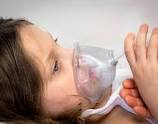
There is no specific treatment available for COVID 19,all over the world.

In such case, the only treatment which can be useful is the symptomatic treatment.

The troublesome symptoms which need to be addressed are ,fever,headache and bodyache apart from cough

The most widely used drug to get relief from fever and pain is paracetamol and acetaminophen derivatives.

Some people do not feel comfortable even after taking Paracetamol and continue to feel headache ,bodyache,and fever.

These cohort of people get relief after taking Ibuprofen.

It was believed at the outset of epidemic of COVID 19,that the use of Ibuprofen may worsen the severity of COVID 19 and people refrained from using this drug.

Researchers from Israel studied the effects of Ibuprofen in patients of COVID 19.They studied 403 patients with the median age of 45 years.They observed the patients from 1 week prior to the diagnosis to,throughout the course of disease.

44% patients developed fever who required its treatment.32% patients used Acetaminophen and 22% used paracetamol to treat their fever.

Respiratory support was needed in  11% patients in Acetaminophen group and approximately10% in Ibuprofen group(P=1).

Mortality was noticed in 2.4% patients using Acetaminophen and 3.8% using Ibuprofen(P=0.95)

  So,there was no difference observed between Acetaminophen group and Ibuprofen group as far as the need for respiratory support or mortality are concerned

Although there are many limitations of this study including recall bias and no study among asymptomatic patients,this is an important study which needs further study on larger population before making a general opinion.

REFERENCES;

Rinott E, Kozer E, Shapira Y, Bar-Haim A, Youngster I. Ibuprofen use and clinical outcomes in COVID-19 patients [published online June, 11 2020]. Clin Microbiol Infect. doi:10.1016/j.cmi.2020.06.003

Certain hand sanitizers are life threatening,Dr.Dev,M.D.,Pediatrician and Pediatric Pulmonologist,Sahibabad,Ghaziabad,Delhi-NCR

In the Pandemic of Coronavirus disease 2019(COVID 19) , hand sanitizers are life saving.

It kills most of the micro-organisms which come in contact with hands.

It should be used when hand washing facility with soap and water is either not available or not feasible.

Hand sanitizers should be composed of ethanol and it should be at least 60% ethanol ,not methanol.

It should be rubbed over hands on all sides till the hands become dry.

Methanol containing hand sanitizers can become dangerous when absorbed through skin or ingested.If there is frequent use of hand sanitizers it can be absorbed through skin.Sometimes it can be ingested by children or inadvertently by adults.Some companies are making and selling methanol containing hand sanitizers.

There has been some reports of death,blindness and hospitalisation after ingestion of methanol containing hand sanitizers by adults and children. Methanol are used to create fuel and antifreeze.

 SIGN AND SYMPTOMS OF METHANOL TOXICITY: Nausea,vomiting,headache,blurred vision.

There may be permanent blindness,seizure,coma,permanent neurological damage and death.

FDA has warned against the use of methanol containing hand sanitizers ,stating that some companies are taking undue advantages of coronavirus pandemic and selling methanol containing hand sanitizers.

It is being recalled from the market .It should be disposed properly using hazardous waste container and should not be poured into drain and should not be mixed with any fluid.

REFERENES;fda.gov

Newborns born to COVID 19 positive mother are not at risk,Dr.Dev,M.D.,Pediatrician and Pediatric Pulmonologist,Sahibabad,Ghaziabad,Delhi-NCR

There is a common belief that infants born to COVID 19 positive mother may have COVID 19,and their immunological status is impaired.

71 Newborns were taken in study to know the facts about the disease status and immunological status.

After exclusion criteria,51 Newborns were included in the study.

All mothers were tested positive for COVID 19 during third trimester of pregnancy.

Chest X-ray of all mothers done before delivery were consistent with viral Pneuimonia.

Apgar scores at 1 minutes and 5 minutes were assessed for all infants.

All Newborns were separated from the mother and kept in isolation with formula feeds.

Caesarean section was the mode of delivery in most cases.

Non of the Newborns showed any symptom in the form of cough ,fever and breathlessness consistent with COVID19, during the course of hospital stay.

No infant required mechanical ventilation but only few needed low flow oxygen with nasal catheter for <3 days.

Blood samples were taken within 3 days of birth to assess immmunological status.

Pharyngeal swabs were taken after 30 minutes of birth to detect Corona virus by RT-PCR .

RT-PCR were negative in all infants.

The levels of lymphocytes subsets,CD3,CD4,CD8 and CD19 and their proportion were normal except for CD16-CD56 which were below normal.

ImmunoglobulinG and Immunuglobulin M were normal.

Interleukin 6 were elevated and it was very high in one Newborn who developed Necrotising enterocolitis in the third week of life.

There was no correlation between duration of COVID 19 in mother and level of lymphocyte subsets or cytokine in infants.

All Newborns were discharged after 3 consequitive samples were found negative for COVID 19.

The study was published in JOURNAL OF ALLERGY AND CLINICAL IMMUNOLOGY.

REFERENCES:Liu P, Zheng J, Yang P, et al. The immunologic status of newborns born to SARS-CoV2-infected mothers in Wuhan, China [published online May 10, 2020]. J Allergy Clin Immunol. doi: 10.1016/j.jaci.2020.04.038

Asthma may be protective against COVID 19,Dr.Dev,M.D.,Pediatrician and Pediatric Pulmonologist,Sahibabad,Ghaziabad,Delhi-NCR

It is a common perception among doctors and public that, asthma may predispose to severe form of Corona Virus Disease 2019(COVID 19).

Corona Virus infects primarily the respiratory tract and Asthma is the most common chronic  disease of  respiratory tract worldwide.

The primary symptoms of Asthma are cough and breathlessness ,which are also the symptoms of Corona Virus Disease 2019.

Asthma is an allergic disease of airway in most of the cases in children ,whereas COVID 19 is an infective disease.

Viruses,chiefly Rhinovirus and Respiratory Syncytial Virus(RSV) are the trigger factors for asthma exacerbation.

So,it is presumed that Corona Virus can also trigger asthma and both may cause serious condition.

Since the airway epithelium and leucocytes of persons suffering from asthma have impaired production of antiviral interferons(Interferon alpha,beta,gamma),due either to allergic inflammations or primarily,it was presumed that the innate immunity of these individuals can not check the viral infections from travelling to lower respiratory tract form upper respiratory tract.

Literature search from 8 studies,comprising more than 17ooo individuals form different geographical areas revealed that the prevalance of COVID 19 was low in areas where prevalance of asthma was high .In persons having comorbidities like Diabetes Mallitus and COPD,Corona Virus infection caused severe infections which was not seen in comorbidities like Asthma.

According to a research,it has been found that 2 host molecules are important for severe acute respiratory syndrome corovirus 2(SARS COV 2).Corona Virus uses ACE 2 receptor (angiotensin converting enzyme 2 receptor) to enter the cell and transmembrane serine protease for priming of S protein  of the virus.

In vitro treatment of epithelial cells with interferons increased the expression of ACE 2 receptor.

The high level of interleukin 13 and T2 cytokines  downregulate the expression of ACE2 in epithelial cells.

Asthmatic individuals have high levels of T2 cytokines and interleukin 13.

Bronchial epithelium of asthmatic individuals have low expression of ACE2 receptor as compared to general population,whereas persons having severe form of COVID 19 show high level of interferon.

Researchers concluded that Asthma may be a protective factor for COVID 19 as asthmatic respiratory epithelium shows low expression of ACE2 receptor.

The study is published in Journal of Allergy and clinical immunology.

REFERENCES:

1.Matsumoto K, Saito H. Does asthma affect morbidity or severity of Covid-19? [published online May 26, 2020]. Editorial. J Allergy Clin Immunol. doi:10.1016/j.jaci.2020.05.017

2. Zheng X-y, Xu Y-j, Guan W-j, Lin L-f. Regional, age and respiratory-secretion-specific prevalence of respiratory viruses associated with asthma exacerbation: a literature review. Arch Virol. 2018;163(4):845-853.

3. Edwards MR, Strong K, Cameron A, Walton RP, Jackson DJ, Johnston SL.Viral infections in allergy and immunology: How allergic inflammation influences viral infections and illness. J Allergy Clin Immunol. 2017;140(4):909-920.

4. Li X, Xu S, Yu M, et al. Risk factors for severity and mortality in adult COVID-19 patients in Wuhan [published online April 12, 2020]. J Allergy Clin Immunol. doi:10.1016/j.jaci.2020.04.006.

5. Singer AJ, Morley EJ, Meyers K, et al. Cohort of 4404 persons under investigation for COVID-19 in a NY hospital and predictors of ICU care and ventilation [published online May 11, 2020]. Ann Emerg Med. doi:10.1016/j.annemergmed.2020.05.011

6. Hoffmann M, Kleine-Weber H, Schroeder S, et al. SARS-CoV-2 cell entry depends on ACE2 and TMPRSS2 and is blocked by a clinically proven protease inhibitor. Cell. 2020;181(2):271-280.e8.


Children display different symptoms in comparision to adults ,of COVID 19,Dr.Dev,M.D.,Pediatrician and Pediatric Pulmonologist,Sahibabad,Delhi-NCR

According to a systematic literature riview,it was found that children have not the same sets of symptoms as adults when they get infected to novel Corona Virus,SARS Cov 2, responsible for Coronavirus disease 2019(COVID19).

The study included clinical,pathological and radiological features.

Researchers included 38 studies comprisisng 1124 cases.They found that about 14 percent children infected with Novel Corona Virus did not exhibit any symptom and they were asymptomatic. About 36 percent were having mild symptoms.,about 46 percent displayed moderate symptoms ,about 2 percent were severly symptomatic while about 1 percent children were critically ill.

The most common symptom was fever which was seen in about 47% of cases,followed by cough which was present in about 41% of cases. Nasal symptoms were seen in about 11% of cases whereas dairrhoea was seen in about 8% cases.

Nausea and vomiting were present in about 7 % of cases and about 37% children developed Pneumonia.About 11% children were diagnosed as upper respiratory tract infections due to Corona Virus.

About 1 in 10 children showed lymphopenia whic is very common in adults.63% children had findings on CECT Chest including ground glass appearance,patchy opacities and consolidation.

There was only 1 death in this systematic review of literature

Fever and cough should not be considered as a hallmark of COVID 19 in children.Pediatrician should keep a high index of suspician to diagnose COVID 19.

Most of the children have favourable outcome after getting infected with novel Corona Virus

child with COVID 19REFERENCES;Pediatrc Pulmonology,online publication,June 2020

Positve test result after recovery from COVID19 is harmless,Dr.Dev,M.D.,Pediatrician and Pediatric Pulmonologist,Sahibabad,Delhi-NCR

It is a cause of worry for many patients including health care workers to receive positive test result for Corona Virus even weeks to months after they have recovered fully.

They are confused and their employers are more confused whether they should join their duties or not.

Mothers are more worried after recovery from disease ,when they should kiss their babies if test report is still positive.

RT-PCR test being used to diagnose Corona Virus Disease 2019 ,detects the pieces of RNA of virus.It can not differentiate between active and dead virus.So,this test is useful for the diagnosis of disease but not for the recovery from the disease.

There is a study of 9 patients suffering from COVID 19,in Germany.They put the sample after 8 days of onset of disease,into the culture.There was no growth of virus in the culture.Moreover,there was no  yield of subgenomic mRNA.These both are indicators of live virus which can infect others and capable of producing disease.It was concluded that if the number of Virus is less than 10000/ml of Sputum,it can not cause the disease..The study was published in NATURE

Resarchers from South Korea,centre for disease control, traced 285 patients who became negative and their isolation was terminated.They again became positive but non of them could transmit the disease to anyone even after converting to positive test result again.

According to the CDC guideline ,a patient is labelled negative only after 2 consequitive test results are negative,keeping in view so many false negative results.

But now it is a fact that some patients may remain positive months after the recovery from COVID 19.

On the other hand,some patients may become positive again after becoming negative.

These patients do not transmit virus that can cause disease to others.

REFERENCES;Doctors Wonder What to Do When Recovered COVID-19 Patients Still Test Positive – Medscape – Jun 09, 2020.