Ozone and Asthma video in Hindi in Children
Uncategorized
Becoming Alive after declared dead-Dr Dev Kumar Jha, Child Specialist & Child chest Super Specialist,L-27,L-Block,Lajpat Nagar,Sahibabad,Delhi NCR
LAZARUS SYNDROME
Visit Dr Dev Child Health Care & Child Chest Super Speciality Clinic ,L-27,Lajpat Nagar,L-Block .Near Kaali Mandir,Sahibabad,Delhi NCR for
All types of Childhood diseases and Super Speciality Treatment for child Chest Diseases
Contact-01203158081,9971720128
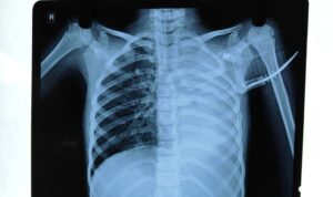
Collapse of Lung,Dr Dev Kumar Jha,M.D. Child specialist and child chest Superspecialist,Delhi and NCR
 This child had been admitted in critical condtion in our Pediatric ICU.
The child was in respiratory distress with decompensated shock.
After initial fluid bolus,vasopressors were started.
The child was maintaining saturation of 96% on room air but at the cost of increased respiratory rate and chest retraction.
The child was intubated and put on mechanical ventilator, to save the respiratory muscles from becoming fatigued.
The child became comfortable with no chest retraction, with saturation of 98% on ventilator.
After few hours,saturation decreased to 70% and ventilator settings were escalated to maintain the saturation.
On chest X-Xray examination, there were complete collapse of left lung which was detected to be responsible for decrease in saturation.
Chest phsiotherapy was started and the repeat chest X-Ray after few hours was normal.
The cause of lung collapse was mucus plug ,obstructiing the left main bronchus which was cleared by chest physiotherapy.
KEY MESSAGE– When the child is on ventilator and saturation falls, percuss and auscultate the chest.
If percussion note is dull and breath sound is diminished on that side ,suspect collapse of lung and do chest X-Ray to detect it.
Apart from mucus plug ,if the Endotracheal tube is placed inadvertantly low so as to enter the right main bronchus, there will be collapse of the left lung and hyperinflation of the right lung.
The tip of the endotracheal tube should lie 1.5 cm above the carina in case of children, which correspond between T2 to T4 vertebrae. It should be checked by chest X-Ray . Neck should be in neutral position,neither extended nor flexed whild doing the chest X-Ray.
On the other hand,if percussion note is hyper-resonant and breath sound is diminished,suspect Pneumothorax.
DR.DEV KUMAR JHA,MBBS(GOLD MEDALIST),M.D.(PEDIATRICS),FELLOWSHIP IN PEDIATRIC PULMONOLOGY,AIIMS,NEW DELHI
USUAL SYMPTOMS,UNUSUAL DIAGNOSIS,A CASE REPORT,Dr.Dev,M.D.,Pediatrician and Pediatric Pulmonologist,Mohan Nagar,Sahibabad,Ghaziabad,Delhi NCR
 11 years old female child presented to our clinic with complaints of
Fever for 24 days,
cough for 15 days
less appetite for 10 days
pain abdomen for 7 days
headache for 5 days
swelling of both lower limbs for 3-4 days
On examination;
Temperature 103 dF,pallor +,icterus+
Pulse rate 56/m regular(relative bradycardia)
Respiratory rate 26/minute ,regular
Generalised lymphadenopathy
bilateral pedal edema
Abdomen-not distended,no tenderness,hepatosplenomegaly+
Chest-bilateral equal breath sound,bilateral vesicular breath sound,bilateral crackles
CVS-NAD
CNS-NAD
Following diagnoses were suspected
Malaria
Typhoid
Viral hepatitis with secondary bacterial infection
Disseminated tuberculosis
Leptospirosis
Scrub typhus
Investigations were planned and done accordingly.
Hb 9 gm/dl
TLC 6800/cmm,P56,L34,E03,M07,B00,Platelet 90000/cmm
SGPT-102,SGOT146,ALP 200,S.BIL 3.8mg/dl
Serum Sodium 130mE/L,Serum potassium 4.4mE/dl
Serum widal-negative
Malaria antigen-negative
Chest X-Ray -bilateral reticulonodular opacities with bilateral pleural effusion
Mantoux test-negative
GA for CBNAAT -negative
The child was emperically treated as enteric fever with cefixime but there was no response.Then the child was treated emperically for malaria and again there was no response.
At this point of time,it was suspected that the child may be suffering from scrub typhus as the child travelled to Uttrakhand during last month.
IgM ELISA for scrub typhus was sent and it came to be positive and the child responded well to doxycycline.
DISCUSSION
:Rickettsia is intracellular ,gram negative proteobacteria with coccobacillar shapes.The disease caused by it is called Rickettsiosis.It is a zoonosis ,transmitted into human by mites(chiggers),ticks or fleas and rodents. Humen are the incidental host.No human to human transmission has been observed.
This disease is frequently seen in Uttrakhand,Rajsthan,assam.West Bengal ,Jammu and Kashmir,Maharashtra Tamil Nadu and Kerala region of India and many countries out of India.
pathogens causing disease in human have been broadly classified into three groups
1.Spotted fever group
2.Typhus group
3.Scrub typhus group
Scrub typhus is causing a health impact in Asia
Pathogenesis;The principle pathogenetic mechanism is vasculitis involving medium and small vessels.It causes increased vascular permeability by disrupting the endothelial tight junctions due to the bacterial load and tumour necrosis factor.Main cause of mortality is pulmonary edema and cerebral edema.
CLINICAL FEATURES OF SCRUB TYPHUS WHICH IS COMMON IN INDIA:
The usual incubation period is 10-12 days with a variability of 6-21 days.Symptoms varies from mild self limiting to severe causing death.
After the period of approximately 5-6 days of bite by mites,there occurs the formation of ESCHAR(shown in the figure) at the site of inoculation of the mite.It is a painless necrotic lesion over skin resembling a cigarette burnt skin surface.The usual sites are groin,axilla,back,neck and other exposed parts of the body.Eschar, if visibl,e makes a clear cut diagnosis of scrub typhus without any investigation.But it is not ususally seen in children.Its prevalence varies from 7-80%.
After about 5-7 days flu like symptoms occurs.In most of the cases fever has been observed with severe headache.
There may be myalgia,weakness,pain abdomen,vomiting,diarrhoea,cough.
O/E There may be relative bradycardia,generalised lymphadenopathy,generalised body rashes and pedal edema
On systemic examination:there may be hepatosplenomegaly
On investigation:
BLOOD-there may be anemia,thrombocytopenia,raised liver enzymes,raised serum bilirubin,hyponatremia,raised blood urea and serum creatinine
Chest X-Ray may show reticulonodular opacities,features of pulmonary edema,bilateral pleural effusion
ECG may shows features of myocarditis with nonspecific ST-T changes,features of heart block
LUMBAR PUNCTURES :CSF pictures are indicative of meningoencephalitis.The CSF picture is similar to Tuberculous meningitis with lymphocytic pleocytosis and raised protein.
DIAGNOSIS; The gold standard is INDIRECT IMMUNOFLUORECENT ANTIBODY TEST(IFA), but it is not available everywhere.The next best is IgM ELISA which is widely available and should be done as the sensitivity of Weil-Felix test is very poor.
TREATMENT.The drug of choice is doxycycline,oral or i.v. in the dose of 2.2 mg/kg 12 hourly below 40 kg of weight and 100 mg b.d. above 40 kg of weight,for a period of 7 days of 3 days after the fever subsides.Now it has been recommended for children of any age to treat Rickettsial diseases as it has not caused enamel hypoplasia or teeth staining even after multiple courses.
Alternative medicine is Azithromycin in the dose of 10 mg/kg/day
Other drugs which may be used in special cases are -clarithromycin.chloramphanicol and Rifampicin
Mortality is above 50% if not recognised and treated timely
COMPLICATIONS.HLH(hemophagocytic lymphohistiocytosis and it is very serious complication.
REFERENCES:
AbdadMY,Abou AbdallahR,FoumierP-E,StenosJ,Vasoo S.Aconcise review of the epidemiology and diagnostics of rickettsioses:Rickettsia and Orientia spp.J clin Microbiol.2018;56:eo1728-17
IssacR,VargheseGM,MathaiE,et al.Scrub typhus:prevalence and diagnostic issue in rural southern India.Clin infect dis.;2004:39;1395-6
Rathi N ,Kulkarni A ,Yewale V;Indian Academy of Pediatrics Guidelines on Rickettsial diseases in children committee.IAP Guideline on Rickettsial disesase in children.Indian Pediatr.2017;54:223-9
Elisabeth BN,Cristina S,DidIer R,Phillipe P.Treatment of Ricketssial spp.infections:a review.Exp rev anti infect Ther.2012;10:1425-37
CORONAVIRUS DISEASE 2019,(COVID 19),Dr.Dev,Pediatric Pulmonologist,Sahibabad,UP,NCR-Delhi
Corona virus is a novel virus,known as severe acute respiratory syndrome virus-2(SARS CoV-2).It was first identified in the Wuhan city of china in 2019,so the disease is termed as COVID 2019.From China ,it has travelled to every part of world making human as a vehicle.It has killed many people world wide.
In 80% of infected people it causes mild disturbances and may go unnoticed.In about 12 % of cases ,infected persons become severe enough to be admitted in hospital and in about 6% cases ,the infected persons need ICU care.Mortality due to this disease is about 2%.
ROUTE OF ENTRANCE INTO THE BODY: It enters into the body through nose ,when an infected person coughs in the near vicinity of an individual.The virus may survive in air,on the surfaces and fomites for a variable period.It also comes in saliva in contrast to common cold virus.So,it may be inhaled from these sites.
The incubation period is 8-37 days,with an average 14 days.An infected person can shed virus for a minimum of 8 days to a maximum of 37 days.
The virus mainly damages the respiratory epithelium and alveoli of lungs causing pneumonia.
CLINICAL FEATURES;Symptoms are cough,sore throat,difficulty in breathing,headache,bodyache,wekness,confusion,dizziness and in some cases,loose motion and vomiting
LABORATORY FINDINGS;Commonb findings on CBC are,leukopenia,lymphopenia and leukocytosis.Other abnormalities are neutrophilia,thrombocytopenia and anemia.
In severe cases,there may be raised D-dimer and prolonged prthrombin time.
IMAGING;Chest X-Ray shows bilateral lung infiltrates in 75% of cases and unilateral lung infiltrates in 25% of cases.
Confirmation of diagnosis; RT-PCR of lower respiratory tract specimen(sputum,tracheal aspirate,BAL fluid) confirms the diagnosis.Sensitivity of upper respiratory tract specimen(throat swab,nasal swab,nasopharyngeal swab )is low.
HIGH RISK OF MORTALITY:Older age,high SOFA score(sequential organ function assessment) and serum level of D-Dimer > 1mcg/ml are the contion with very high mortality.
TREATMENT;There is no definite treatment as of now.Some antivirals have been tried like oseltamavir,ritonavir and lopinavir with variable results.Hydroxychloroquine has also been tried.
PREVENTION;Prevention is the only cure.Maintain respiratory etiquette,avoid crowded places,and maintain a distance of at least 1 meter(3 feets)
REFERENCES: 1.BMJ Best practice COVID 19
2.Lancet.doi:10.1016/SO140-6736(20)30566-3
Your website’s article title 05 edit by going to dashboard

Lorem ipsum dolor sit amet, consectetur adipiscing elit. Integer nec odio. Praesent libero. Sed cursus ante dapibus diam. Sed nisi. Nulla quis sem at nibh elementum imperdiet. Duis sagittis ipsum. Praesent mauris. Fusce nec tellus sed augue semper porta. Mauris massa. Vestibulum lacinia arcu eget nulla. Class aptent taciti sociosqu ad litora torquent per conubia nostra, per inceptos himenaeos.
Curabitur sodales ligula in libero. Sed dignissim lacinia nunc. Curabitur tortor. Pellentesque nibh. Aenean quam. In scelerisque sem at dolor. Maecenas mattis. Sed convallis tristique sem. Proin ut ligula vel nunc egestas porttitor. Morbi lectus risus, iaculis vel, suscipit quis, luctus non, massa. Fusce ac turpis quis ligula lacinia aliquet. Mauris ipsum. Nulla metus metus, ullamcorper vel, tincidunt sed, euismod in, nibh.
Quisque volutpat condimentum velit. Class aptent taciti sociosqu ad litora torquent per conubia nostra, per inceptos himenaeos. Nam nec ante. Sed lacinia, urna non tincidunt mattis, tortor neque adipiscing diam, a cursus ipsum ante quis turpis. Nulla facilisi. Ut fringilla. Suspendisse potenti. Nunc feugiat mi a tellus consequat imperdiet. Vestibulum sapien. Proin quam. Etiam ultrices. Suspendisse in justo eu magna luctus suscipit. Sed lectus. Integer euismod lacus luctus magna.
Vestibulum lacinia arcu
Quisque cursus, metus vitae pharetra auctor, sem massa mattis sem, at interdum magna augue eget diam. Vestibulum ante ipsum primis in faucibus orci luctus et ultrices posuere cubilia Curae; Morbi lacinia molestie dui. Praesent blandit dolor. Sed non quam. In vel mi sit amet augue congue elementum. Morbi in ipsum sit amet pede facilisis laoreet. Donec lacus nunc, viverra nec, blandit vel, egestas et, augue. Vestibulum tincidunt malesuada tellus. Ut ultrices ultrices enim. Curabitur sit amet mauris. Morbi in dui quis est pulvinar ullamcorper. Nulla facilisi.
Integer lacinia sollicitudin massa. Cras metus. Sed aliquet risus a tortor. Integer id quam. Morbi mi. Quisque nisl felis, venenatis tristique, dignissim in, ultrices sit amet, augue. Proin sodales libero eget ante.
Aenean lectus elit, fermentum non, convallis id, sagittis at, neque. Nullam mauris orci, aliquet et, iaculis et, viverra vitae, ligula. Nulla ut felis in purus aliquam imperdiet. Maecenas aliquet mollis lectus. Vivamus consectetuer risus et tortor. Lorem ipsum dolor sit amet, consectetur adipiscing elit. Integer nec odio. Praesent libero. Sed cursus ante dapibus diam. Sed nisi. Nulla quis sem at nibh elementum imperdiet. Duis sagittis ipsum.
Hello world!
Welcome to WordPress. This is your first post. Edit or delete it, then start writing!