ABSTRACT:
Tropical Pulmonary eosinophilia is a rare disease affecting multiple organs but there is more involvement of lungs.It ia caused by hypersensitivity reactions to trapped microfilariae of Wuchereria bancrofti(W.bancrofti) and Brugia malayi(B.malayi) in the lungs. I report a case of TPE(Tropical Pulmonary eosinophilia in a child who presented similar to Pulmonary tuberculosis,diagnosed and treated timely with good response.
KEYWORDS Tropical pulmonary eosinophilia,hypersesitivity reactions, mocrofilaria,Filaria
INTRODUCTION:
Tropical Pulmonary eosinophilia as a disease entity was first described in 1940 and the term Pseudotuberculosis with eosinophilia was given.In 1943 Weingarten coined the term TPE(Tropical Pulmonary eosinophilia) for this disease. The disease occurs in costal India like Maharashtra,Kerala,Goa and odisha.It has also been found in some areas of Bihar( prevalence of 9% in jail inmates of Patna).A prevalence of 0.5% among children of Tamil Nadu has been found.It occurs in filaria endemic zones of South America,Africa and South East Asia.The common presentation is cough,difficulty in breathing,fever and weight loss. On investigation there is Leucocytosis with eosinophilia,raised serum IgE and raised specific serum IgE and IgG against Filaria antigen. Treatment is 3-4 weeks course of DEC(Diethylecarbamzepine)
CASE SUMMARY:
9 years old female child came to my OPD with complaint of fever off and on for 2-3 months,cough for 2 months,difficulty in breathing for 1 month ,breathless on exertion for 1 month.
There was no contact history for tuberculosis in the family or relatives
She was taking treatment from different doctors in past 3 months
She was also loosing her weight in last 2 months and she was easily fatigued in last 1 month
on clinical examination she was underweight and anemic,there was significant cervical lymphadenopathy ,no clubbing,no cyanosis and on chest auscultation there were bilateral wheezes and crackles.Abdominal examination showed hepatsplenomegaly
On investigation
Hb 14.3g/dl,
TLC-50,000/cmm,
Neutrophil 28%,lymphocytes10%,
Eosinophil 61%
Basophil 0%,ESR18mm/1H,
Peripheral smear examination-leucocytes increased in number with normal distribution, platelets adequate,no blast cells, malaria parasites amd microfilariae absent
Chest X-Ray-multiple bilateral miliary mottling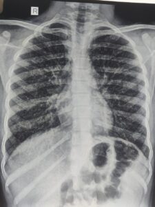
I thought it may be a case of
1.Tuberculosis with some parasitic infections,
2.Asthma with ABPA,
3.hypereosinophilic syndrome,
4.Tropical Pulmonary Eosinophilia
Then , I investigated further
GA for CBNAAT -Negative,Montoux test -negative
Serum IgE -3389U/L,Serum specific filaria antibody IgE and Ig G were raised
Then,I Put the child on DEC 5 mg/kg/day for 4 weeks orally and child started improving after 1 week of initiating DEC
Repeat Serum IgE after 4 weeks was normal ,TLC came down to normal range, and repeat chest X-Ray at that time was normal,child started gaining weight
DISCUSSION:
TPE is usually seen between the age group of 15-45 years .It is rare to see in children less than 15 years ,which was our case. The male:female is 4;1 which is against our case.The clinical features are malaise,fatigue,cough ,chest pain, breathlessness which are seen more during night.Clinical findings in chest may be wheezes and crackes but in 20% cases,there may be no findings in chest.It may involve any system apart from chest leading to the findings of cervical lymphadenopathy,hepatomegaly,splenomegaly,hepatosplenomegaly
The pathology involves hypersensitivity reactions type I,III,and IV to microfilariae.It is due to release of microfilariae from lymphatics which get entrapped into pulmonary circulation.Degranulation of eosinophils, release major basic proteins along with other granules which destruct microfilariae as well as respiratory and terminal bronchioles with lung parenchyma.In long standing untreated cases fibrosis develops.
Lung function test shows mixed obstructive and restrictive pattern of lung disease.
Chest radiograph shows raticuonodular opacities in mid zone and lower zone but as the disease progresses,there are miliary mottling all over the lung fields. Chest X-Ray may be normal in about 20% cases.CT chest may show mediastinal lymphadenopathy,consolidation,bronchiectasis,cavitation and Pleural effision.
CBC shows leococytosis with marked eosinophilia,AEC is usually more than 3000/cmm,Total serum IgE and specific IgE and IgG against Filaria is raised.
ELISA test detecting 0g4c3 antigen is sensitive and specific to W.bancrofti and Sanwich ELISA detecting recombinant antigen Bm-SXP-1 is specific to B.malayi
Trearment is DEC,5mg/kg/day for 4 weeks.Steroids may have a role but dose and duration is yet to be known
Our case is well fitting into the diagnostic criteria
CONCLUSION: TPE is a hypersensitive reaction to W.Bancrofti and B.malayi microfilariae mainly type I, causing eosinophilic alveolitis with inflammation of terminal an d respiratory bronchioles.Diagnosed by clinical features ,mainly night symptoms of cough and dyspnea,peripheral eosinophilia which has diurnal variation and paradoxically low level during night,raised serum IgE and specific IgE and IgG against filaria ,infiltrates on chest radiograph.Treatment is oral DEC for 4 weeks but there is 20% relapse within 5 years. The disease closely resembles Pulmonary tuberculosis and asthma.Untreated and partialy treated case may progress to Interstitial lung disease(ILD)
REFERENCES:
1.Indian J Med Res. 2013 Sep; 138(3): 295–302.
2. Udwadia FE. Herzog H, editor. Tropical eosinophilia. Pulmonary eosinophilia: progress in pulmonary research. 1975;VII:35–155. Basel: S Karger
3.Neva FA, Kaplan AP, Pacheco G, Gray L, Danaraj TJ. Tropical eosinophilia. A human model of parasitic immunopathology, with observations on serum IgE levels before and after treatment. J Allergy Clin Immunol. 1975;55:422–9
4. Frimodt-Moller C, Barton RM. A pseudo-tuberculosis condition associated with eosinophilia. Indian Med Gaz. 1940;75:607–13