ABSTRACT;
Necrotising pneumonia is a serious and uncommon complication of pneumonia in children.It usually occurs in previously healthy children.It should be suspected when a child with pneumonia worsens clinically after 72 hours of appropriate antibiotics. It may be complicated by pyopneumothorax and Broncopleural Fistula. It is diagnosed by radioimaging of chest and CECT Chest is the best modality.It needs long term antibiotics with long hospital stay.
CASE SUMMARY:
8 years male child came in our emergency with history of fever for 4 days with weakness and breathing difficulty for 2 days
On reaching hospital ,in the emergency department the child lost his consciousness
On examination ,the child was in shock and breath sound was diminished on right side of chest
The child had to put on ventilator immediately and then chest X-Ray was done which revealed right sided pneumothorax
Chest tube drainage was done immediately,which relieved pneumothorax
Appropriate antibiotics were started but the condition of the child did not improve
Pus started coming out in the chest tube
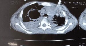
CECT Chest was done which revealed cavitary lesions (thin walled cavities) with distortion of lung architecture with pyopneumothorax
There were appearance of air bubble in the chest tube which was not earlier after relieving of pneumothorax
It was then confirmed that there has been formation of Bronchopleural Fistula(BPF)
In view of necrotising Pneumonia and BPF, antibiotics was upgraded to clindamycin and Meropenem
Ventilator settings was adjusted with low Ti. Low PEEP and minimum Tidal Volume,

 en
en
General condition improved gradually and child became afebrile,OG tube feeding was started while on ventilator
Condition became permissible to extubate the child after 2 weeks of mechanical ventilation
While on ventilator ,the child had persistent metabolic alkalosis after the first report on ABG of respiratory acidosis before ventilation,there was persistent hypokalemia ,then one episode of respiratory alkalosis,all were managed properly. Child required one PRBC transfusion while on ventilator.
Chest tube could be removed after 3 weeks as BPF healed on conservative management
Now the child is on oral antibiotics and doing well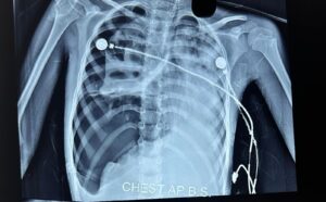

On laboratory investigations- There was leukocytosis,anemia,hypoalbuminemia,raised LDH,raised CRP-123mg/dl
Chest X-Ray showed right sided pneumothorax with multiple cavities.
Chest CT showed Pyonneumothorax with thin walled cavity in right lung.
DISCUSSION:
Necrotising pneumonia is a rare complication of pneumonia in children.
It is being recognised now a days due to increased use of CT scan.
It occurs in previusly healthy children due to high virulence of infecting organism.
Interaction between bacteria and viruses also play a role. The most common responsible organisma are Pneumococcus and Staphylococcus. It may be caused by Mycoplasma and Mycobacterium Tuberculosis.
PVL-Panton valentine leucocidin, secreted by MRSA is responsible in some cases. In such cases leucopenia rather than leucocytosis occurs with fast detrioration in clinical condition.
After infectin,vasculitis occurs in pulmonary vessels due to release of cytokines in which interleukin 8 driven neutrophils at inflammatory site,play an important role,leading to thrombosis,occlusion of vessels,activation of coagulation pathway, which causes liquefaction and necrosis, destruction of lung parenchyma takes place, and there occurs formation of multiple thin walled cysts. Somtimes gangrene occurs in whole lobe of lung.If the necrosis is near pleura,repture of pleura happens and pneumothorax develops. Pus then gets collected into pleural cavity and pyopneumothorax develops. This is the stage of Bronchopleural fistula. Bronchopleural fistula is usually a complication of chest tube drainage or surgical intervention but rarely occurs as an extension of disease itself.
TREATMENT:
It is mainly conservative with intravenous antibiotics with supportive care.Mechanical ventilation is required in case of respiratory failure. Chest tube insertion is avoided initially but situation of tension pneumothorax compells for chest tube drainage sometimes. .Fever persists for a long time. The duration of antibiotics may vary from 2- 6 weeks. I.V antibiotics should be given till the child is afebrile for at least 24 hours, imflammatory markers are coming down, hemodynamics are stable an oral feeds are tolerated.After that oral antibiotics may be needed for further 2- 3 weeks. Some children may require surgical intervention(VATS) to evacuate pus,sealing of Bronchopleural Fistula with FIBRIN GLUE or muscle flap. In some circumstances segmental resection of lung,lobectomy and even pneumenectomy may be required to save the child.
CONCLUSION:
Necrotising pneumonia is a very serious condtion ,requires intensive care ,sometimes with mechanical ventilation,chest tube drainage with fibrinolytics but mortality is very low with adequate treatment. Some cases may require prolonged antibiotics course with average duration of hospital of 28 days. In rare circumstances Video assistaed Thoracoscopic surgery is required.
Keywords:Necrotising pneumonia(NP),Pyopneumothorax,Tension pneumothorax,Bronchopleural Fistula(BPF),Thin walled lung cavity
REFERENCES:
1.Ramgopal S, Ivan Y, Medsinge A, Saladino RA. Pediatric necrotizing pneumonia and review of the literature. Pediatr Emerg Care. 2017;33:112–115. [PubMed] [Google Scholar]
2.McCarthy VP, Patamasucon P, Gaines T, Lucas MA. Necrotizing pneumonia in childhood. Pediatr Pulmonol. 1999;28:217–221. doi: 10.1002/(SICI)1099-0496(199909)28:3<217::AID-PPUL9>3.0.CO;2-R. [PubMed]
3.Gillet Y, Issartel B, Vanhems P, Fournet J-C, Lina G, Bes M, et al. Association between Staphylococcus aureus strains carrying gene for Panton-valentine leucocidin and highly lethal necrotising pneumonia in young immunocompetent patients. Lancet. 2002;359:753–759. doi: 10.1016/S0140-6736(02)07877-7. [PubMed] [CrossRef] [Google Scholar]
4.Kalaskar AS, Heresi GP, Wanger A, Murphy JR, Wootton SH. Severe necrotizing pneumonia in children, Houston, Texas, USA. Emerg Infect Dis. 2009;15:1696–1698. doi: 10.3201/eid1510.090589. [PMC free article] [PubMed] [CrossRef] [Google Scholar]